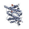
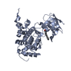
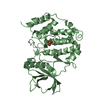
[English] 日本語
Yorodumi
- PDB-2v9t: Complex between the second LRR domain of Slit2 and The first Ig d... -

+
Open data


ID or keywords:

Loading...

-
Basic information

Entry
Database: PDB / ID: 2v9t
TitleComplex between the second LRR domain of Slit2 and The first Ig domain from Robo1
Components
  • ROUNDABOUT HOMOLOG 1
  • SLIT HOMOLOG 2 PROTEIN N-PRODUCT
KeywordsSTRUCTURAL PROTEIN/RECEPTOR / STRUCTURAL PROTEIN-RECEPTOR COMPLEX / DEVELOPMENTAL PROTEIN / IG DOMAIN / ROUNDABOUT / CHEMOTAXIS / LRR DOMAIN / IMMUNOGLOBULIN DOMAIN / LEUCINE-RICH REPEAT / NEURONAL DEVELOPMENT / PHOSPHORYLATION / EGF-LIKE DOMAIN / DISEASE MUTATION / SLIT2 / LIGAND / MEMBRANE / SECRETED / RECEPTOR / NEUROGENESIS / GLYCOPROTEIN / TRANSMEMBRANE / PROTO-ONCOGENE / DIFFERENTIATION STRUCTURAL PROTEIN/RECEPTOR / COMPLEX
Function / homology
Function and homology information


negative regulation of leukocyte chemotaxis / corticospinal neuron axon guidance through spinal cord / negative regulation of mononuclear cell migration / negative regulation of lamellipodium assembly / induction of negative chemotaxis / negative regulation of retinal ganglion cell axon guidance / apoptotic process involved in luteolysis / negative regulation of monocyte chemotaxis / chemorepulsion involved in postnatal olfactory bulb interneuron migration / Roundabout binding ...negative regulation of leukocyte chemotaxis / corticospinal neuron axon guidance through spinal cord / negative regulation of mononuclear cell migration / negative regulation of lamellipodium assembly / induction of negative chemotaxis / negative regulation of retinal ganglion cell axon guidance / apoptotic process involved in luteolysis / negative regulation of monocyte chemotaxis / chemorepulsion involved in postnatal olfactory bulb interneuron migration / Roundabout binding / negative regulation of negative chemotaxis / negative regulation of smooth muscle cell chemotaxis / negative regulation of cellular response to growth factor stimulus / Regulation of cortical dendrite branching / cellular response to heparin / negative regulation of small GTPase mediated signal transduction / negative regulation of mammary gland epithelial cell proliferation / LRR domain binding / negative regulation of chemokine-mediated signaling pathway / response to cortisol / laminin-1 binding / negative regulation of smooth muscle cell migration / heart induction / axon extension involved in axon guidance / axon guidance receptor activity / positive regulation of vascular endothelial growth factor signaling pathway / Netrin-1 signaling / GTPase inhibitor activity / Regulation of commissural axon pathfinding by SLIT and ROBO / Role of ABL in ROBO-SLIT signaling / Inactivation of CDC42 and RAC1 / negative regulation of neutrophil chemotaxis / SLIT2:ROBO1 increases RHOA activity / Roundabout signaling pathway / endocardial cushion formation / negative regulation of actin filament polymerization / Signaling by ROBO receptors / pulmonary valve morphogenesis / outflow tract septum morphogenesis / negative regulation of vascular permeability / motor neuron axon guidance / cell migration involved in sprouting angiogenesis / aortic valve morphogenesis / retinal ganglion cell axon guidance / Activation of RAC1 / positive regulation of vascular endothelial growth factor receptor signaling pathway / axon midline choice point recognition / aorta development / ureteric bud development / proteoglycan binding / negative regulation of endothelial cell migration / positive regulation of Rho protein signal transduction / negative chemotaxis / positive regulation of axonogenesis / ventricular septum morphogenesis / branching morphogenesis of an epithelial tube / positive regulation of Notch signaling pathway / homophilic cell adhesion via plasma membrane adhesion molecules / cellular response to hormone stimulus / endoplasmic reticulum-Golgi intermediate compartment membrane / negative regulation of cell migration / negative regulation of protein phosphorylation / axon guidance / positive regulation of MAP kinase activity / negative regulation of cell growth / Regulation of expression of SLITs and ROBOs / activation of cysteine-type endopeptidase activity involved in apoptotic process / heparin binding / nervous system development / cell adhesion / neuron projection / positive regulation of apoptotic process / axon / negative regulation of gene expression / calcium ion binding / positive regulation of gene expression / cell surface / protein homodimerization activity / extracellular space / extracellular exosome / extracellular region / membrane / identical protein binding / plasma membrane / cytoplasm
Similarity search - Function
Roundabout homologue 1 / Leucine rich repeat C-terminal domain / Follistatin-like, N-terminal / Follistatin-N-terminal domain-like / Laminin G domain / EGF-like, conserved site / Human growth factor-like EGF / C-terminal cystine knot signature. / C-terminal cystine knot domain profile. / Leucine rich repeat N-terminal domain ...Roundabout homologue 1 / Leucine rich repeat C-terminal domain / Follistatin-like, N-terminal / Follistatin-N-terminal domain-like / Laminin G domain / EGF-like, conserved site / Human growth factor-like EGF / C-terminal cystine knot signature. / C-terminal cystine knot domain profile. / Leucine rich repeat N-terminal domain / Cystine knot, C-terminal / C-terminal cystine knot-like domain (CTCK) / Laminin G domain profile. / Leucine-rich repeat N-terminal domain / Leucine rich repeat N-terminal domain / Laminin G domain / Laminin G domain / Leucine Rich Repeat / Cysteine-rich flanking region, C-terminal / Leucine rich repeat C-terminal domain / Leucine-rich repeat, LRR (right-handed beta-alpha superhelix) / Ribonuclease Inhibitor / Leucine-rich repeat, SDS22-like subfamily / Alpha-Beta Horseshoe / EGF-type aspartate/asparagine hydroxylation site / EGF-like domain / EGF-like calcium-binding, conserved site / Calcium-binding EGF-like domain signature. / Immunoglobulin domain / Aspartic acid and asparagine hydroxylation site. / EGF-like calcium-binding domain / Calcium-binding EGF-like domain / Leucine rich repeat / Leucine-rich repeat, typical subtype / Leucine-rich repeats, typical (most populated) subfamily / Immunoglobulin I-set / Immunoglobulin I-set domain / Epidermal growth factor-like domain. / Leucine-rich repeat profile. / EGF-like domain profile. / EGF-like domain signature 2. / EGF-like domain signature 1. / Leucine-rich repeat / Fibronectin type III domain / EGF-like domain / Fibronectin type 3 domain / Immunoglobulin subtype 2 / Immunoglobulin C-2 Type / Immunoglobulin V-Type / Fibronectin type-III domain profile. / Leucine-rich repeat domain superfamily / Fibronectin type III / Fibronectin type III superfamily / Immunoglobulin V-set domain / Concanavalin A-like lectin/glucanase domain superfamily / Immunoglobulin subtype / Immunoglobulin / Ig-like domain profile. / Immunoglobulin-like domain / Immunoglobulin-like domain superfamily / Immunoglobulins / Immunoglobulin-like fold / Immunoglobulin-like / Sandwich / Mainly Beta / Alpha Beta
Similarity search - Domain/homology
Slit homolog 2 protein / Roundabout homolog 1
Similarity search - Component
Biological speciesHOMO SAPIENS (human)
MethodX-RAY DIFFRACTION / SYNCHROTRON / MOLECULAR REPLACEMENT / Resolution: 1.7 Å
AuthorsMorlot, C. / Cusack, S. / McCarthy, A.A.
Citation
#1: Journal: Acta Crystallogr.,Sect.F / Year: 2007
Title: Cloning, Expression, Crystallization and Preliminary X-Ray Analysis of the First Two Ig Domains from Human Roundabout 1 (Robo1).
Authors: Morlot, C. / Hemrika, W. / Romijn, R.A. / Gros, P. / Cusack, S. / Mccarthy, A.A.
History
DepositionAug 25, 2007Deposition site: PDBE / Processing site: PDBE
Revision 1.0Sep 25, 2007Provider: repository / Type: Initial release
Revision 1.1Jul 13, 2011Group: Advisory / Version format compliance
Revision 1.2Aug 29, 2012Group: Database references
Remark 700 SHEET THE SHEET STRUCTURE OF THIS MOLECULE IS BIFURCATED. IN ORDER TO REPRESENT THIS FEATURE IN ... SHEET THE SHEET STRUCTURE OF THIS MOLECULE IS BIFURCATED. IN ORDER TO REPRESENT THIS FEATURE IN THE SHEET RECORDS BELOW, TWO SHEETS ARE DEFINED.

-
Structure visualization

Structure viewerMolecule:
MolmilJmol/JSmol

Downloads & links

-
Assembly

Deposited unit
A: ROUNDABOUT HOMOLOG 1
B: SLIT HOMOLOG 2 PROTEIN N-PRODUCT


Theoretical massNumber of molelcules
Total (without water)37,7802
Polymers37,7802
Non-polymers00
Water6,936385
1


  • Idetical with deposited unit
  • defined by author&software
TypeNameSymmetry operationNumber
identity operation1_555x,y,z1
Buried area1560 Å2
ΔGint-6.2 kcal/mol
Surface area18750 Å2
MethodPQS
Unit cell
Length a, b, c (Å)51.270, 190.090, 40.870
Angle α, β, γ (deg.)90.00, 90.00, 90.00
Int Tables number18
Space group name H-MP21212

-
Components

#1: Protein ROUNDABOUT HOMOLOG 1 / H-ROBO-1 / DELETED IN U TWENTY TWENTY / ROBO1


Mass: 13103.687 Da / Num. of mol.: 1 / Fragment: IG1-2M, RESIDUES 61-166 / Mutation: YES
Source method: isolated from a genetically manipulated source
Source: (gene. exp.) HOMO SAPIENS (human) / Cell line (production host): HEK293-EBNA1 / Production host: HOMO SAPIENS (human) / References: UniProt: Q9Y6N7
#2: Protein SLIT HOMOLOG 2 PROTEIN N-PRODUCT / SLIT-2 / SLIT2 / SLIT HOMOLOG 2 PROTEIN


Mass: 24676.551 Da / Num. of mol.: 1 / Fragment: SECOND LRR DOMAIN, RESIDUES 271-479
Source method: isolated from a genetically manipulated source
Source: (gene. exp.) HOMO SAPIENS (human) / Cell line (production host): HEK293-EBNA1 / Production host: HOMO SAPIENS (human) / References: UniProt: O94813
#3: Water ChemComp-HOH / water / Water


Mass: 18.015 Da / Num. of mol.: 385 / Source method: isolated from a natural source / Formula: H2O
Compound detailsENGINEERED RESIDUE IN CHAIN A, ASN 160 TO ASP
Sequence detailsA N160D MUTATION WAS MADE TO PREVENT POTENTIAL GLYCOSYLATION

-
Experimental details

-
Experiment

ExperimentMethod: X-RAY DIFFRACTION / Number of used crystals: 1

-
Sample preparation

CrystalDensity Matthews: 2.6 Å3/Da / Density % sol: 53 % / Description: NONE
Crystal growpH: 5.6 / Details: 12% PEG 6000, 0.1M LI SO4, 0.1M NA CITRATE, PH=5.6

-
Data collection

DiffractionMean temperature: 100 K
Diffraction sourceSource: SYNCHROTRON / Site: ESRF / Beamline: ID23-1 / Wavelength: 0.9537
DetectorType: ADSC CCD / Detector: CCD / Date: Jan 26, 2007 / Details: TORODIAL MIRROR
RadiationMonochromator: SI(111) / Protocol: SINGLE WAVELENGTH / Monochromatic (M) / Laue (L): M / Scattering type: x-ray
Radiation wavelengthWavelength: 0.9537 Å / Relative weight: 1
ReflectionResolution: 1.7→30 Å / Num. obs: 42371 / % possible obs: 98.1 % / Observed criterion σ(I): 1 / Redundancy: 3.8 % / Rmerge(I) obs: 0.08 / Net I/σ(I): 12.9
Reflection shellResolution: 1.7→1.8 Å / Redundancy: 3.7 % / Rmerge(I) obs: 0.53 / Mean I/σ(I) obs: 2.5 / % possible all: 94.9

-
Processing

Software
NameVersionClassification
REFMAC5.2.0019refinement
XDSdata reduction
XSCALEdata scaling
PHASERphasing
RefinementMethod to determine structure: MOLECULAR REPLACEMENT / Resolution: 1.7→19.98 Å / Cor.coef. Fo:Fc: 0.956 / Cor.coef. Fo:Fc free: 0.938 / SU B: 4.559 / SU ML: 0.077 / TLS residual ADP flag: LIKELY RESIDUAL / Cross valid method: THROUGHOUT / ESU R: 0.1 / ESU R Free: 0.103 / Stereochemistry target values: MAXIMUM LIKELIHOOD / Details: HYDROGENS HAVE BEEN ADDED IN THE RIDING POSITIONS.
RfactorNum. reflection% reflectionSelection details
Rfree0.226 2234 5.1 %RANDOM
Rwork0.186 ---
obs0.188 41951 98.2 %-
Solvent computationIon probe radii: 0.8 Å / Shrinkage radii: 0.8 Å / VDW probe radii: 1.2 Å / Solvent model: MASK
Displacement parametersBiso mean: 22.96 Å2
Baniso -1Baniso -2Baniso -3
1--0.07 Å20 Å20 Å2
2---0.02 Å20 Å2
3---0.09 Å2
Refinement stepCycle: LAST / Resolution: 1.7→19.98 Å
ProteinNucleic acidLigandSolventTotal
Num. atoms2473 0 0 385 2858
Refine LS restraints
Refine-IDTypeDev idealDev ideal targetNumber
X-RAY DIFFRACTIONr_bond_refined_d0.0150.0222557
X-RAY DIFFRACTIONr_bond_other_d
X-RAY DIFFRACTIONr_angle_refined_deg1.5651.9813482
X-RAY DIFFRACTIONr_angle_other_deg
X-RAY DIFFRACTIONr_dihedral_angle_1_deg6.5785326
X-RAY DIFFRACTIONr_dihedral_angle_2_deg40.89223.784111
X-RAY DIFFRACTIONr_dihedral_angle_3_deg13.77815447
X-RAY DIFFRACTIONr_dihedral_angle_4_deg19.0341520
X-RAY DIFFRACTIONr_chiral_restr0.1120.2400
X-RAY DIFFRACTIONr_gen_planes_refined0.0060.021923
X-RAY DIFFRACTIONr_gen_planes_other
X-RAY DIFFRACTIONr_nbd_refined0.2170.21215
X-RAY DIFFRACTIONr_nbd_other
X-RAY DIFFRACTIONr_nbtor_refined0.3130.21715
X-RAY DIFFRACTIONr_nbtor_other
X-RAY DIFFRACTIONr_xyhbond_nbd_refined0.1670.2327
X-RAY DIFFRACTIONr_xyhbond_nbd_other
X-RAY DIFFRACTIONr_metal_ion_refined
X-RAY DIFFRACTIONr_metal_ion_other
X-RAY DIFFRACTIONr_symmetry_vdw_refined0.2680.238
X-RAY DIFFRACTIONr_symmetry_vdw_other
X-RAY DIFFRACTIONr_symmetry_hbond_refined0.1680.212
X-RAY DIFFRACTIONr_symmetry_hbond_other
X-RAY DIFFRACTIONr_symmetry_metal_ion_refined
X-RAY DIFFRACTIONr_symmetry_metal_ion_other
X-RAY DIFFRACTIONr_mcbond_it1.0541.51657
X-RAY DIFFRACTIONr_mcbond_other
X-RAY DIFFRACTIONr_mcangle_it1.58322605
X-RAY DIFFRACTIONr_mcangle_other
X-RAY DIFFRACTIONr_scbond_it2.69831003
X-RAY DIFFRACTIONr_scbond_other
X-RAY DIFFRACTIONr_scangle_it4.4394.5871
X-RAY DIFFRACTIONr_scangle_other
X-RAY DIFFRACTIONr_long_range_B_refined
X-RAY DIFFRACTIONr_long_range_B_other
X-RAY DIFFRACTIONr_rigid_bond_restr
X-RAY DIFFRACTIONr_sphericity_free
X-RAY DIFFRACTIONr_sphericity_bonded
LS refinement shellResolution: 1.7→1.74 Å / Total num. of bins used: 20 /
RfactorNum. reflection
Rfree0.335 150
Rwork0.295 2888
Refinement TLS params.

Method: refined / Refine-ID: X-RAY DIFFRACTION

IDL112)L122)L132)L222)L232)L332)S11 (Å °)S12 (Å °)S13 (Å °)S21 (Å °)S22 (Å °)S23 (Å °)S31 (Å °)S32 (Å °)S33 (Å °)T112)T122)T132)T222)T232)T332)Origin x (Å)Origin y (Å)Origin z (Å)
11.0842-0.54290.26431.2204-1.55992.86720.0164-0.05710.20810.0008-0.0151-0.009-0.02470.0985-0.0013-0.0423-0.0081-0.0016-0.021-0.006-0.050311.6691-32.2634-6.7742
21.12940.1587-0.01091.24540.13520.5361-0.006-0.01090.2307-0.0185-0.01590.1357-0.0638-0.05950.0219-0.04740.0088-0.0045-0.02760.0181-0.0181-7.3315-26.9891-16.1992
Refinement TLS group
IDRefine-IDRefine TLS-IDAuth asym-IDAuth seq-ID
1X-RAY DIFFRACTION1A60 - 166
2X-RAY DIFFRACTION2B269 - 478

+
About Yorodumi

-
News

-
Feb 9, 2022. New format data for meta-information of EMDB entries

New format data for meta-information of EMDB entries

  • Version 3 of the EMDB header file is now the official format.
  • The previous official version 1.9 will be removed from the archive.

Related info.:EMDB header

External links:wwPDB to switch to version 3 of the EMDB data model

-
Aug 12, 2020. Covid-19 info

Covid-19 info

URL: https://pdbj.org/emnavi/covid19.php

New page: Covid-19 featured information page in EM Navigator.

Related info.:Covid-19 info / Mar 5, 2020. Novel coronavirus structure data

+
Mar 5, 2020. Novel coronavirus structure data

Novel coronavirus structure data

Related info.:Yorodumi Speices / Aug 12, 2020. Covid-19 info

External links:COVID-19 featured content - PDBj / Molecule of the Month (242):Coronavirus Proteases

+
Jan 31, 2019. EMDB accession codes are about to change! (news from PDBe EMDB page)

EMDB accession codes are about to change! (news from PDBe EMDB page)

  • The allocation of 4 digits for EMDB accession codes will soon come to an end. Whilst these codes will remain in use, new EMDB accession codes will include an additional digit and will expand incrementally as the available range of codes is exhausted. The current 4-digit format prefixed with “EMD-” (i.e. EMD-XXXX) will advance to a 5-digit format (i.e. EMD-XXXXX), and so on. It is currently estimated that the 4-digit codes will be depleted around Spring 2019, at which point the 5-digit format will come into force.
  • The EM Navigator/Yorodumi systems omit the EMD- prefix.

Related info.:Q: What is EMD? / ID/Accession-code notation in Yorodumi/EM Navigator

External links:EMDB Accession Codes are Changing Soon! / Contact to PDBj

+
Jul 12, 2017. Major update of PDB

Major update of PDB

  • wwPDB released updated PDB data conforming to the new PDBx/mmCIF dictionary.
  • This is a major update changing the version number from 4 to 5, and with Remediation, in which all the entries are updated.
  • In this update, many items about electron microscopy experimental information are reorganized (e.g. em_software).
  • Now, EM Navigator and Yorodumi are based on the updated data.

External links:wwPDB Remediation / Enriched Model Files Conforming to OneDep Data Standards Now Available in the PDB FTP Archive

-
Yorodumi

Thousand views of thousand structures

  • Yorodumi is a browser for structure data from EMDB, PDB, SASBDB, etc.
  • This page is also the successor to EM Navigator detail page, and also detail information page/front-end page for Omokage search.
  • The word "yorodu" (or yorozu) is an old Japanese word meaning "ten thousand". "mi" (miru) is to see.

Related info.:EMDB / PDB / SASBDB / Comparison of 3 databanks / Yorodumi Search / Aug 31, 2016. New EM Navigator & Yorodumi / Yorodumi Papers / Jmol/JSmol / Function and homology information / Changes in new EM Navigator and Yorodumi

Read more