 Movie
Movie Controller

[English] 日本語
 Yorodumi
- PDB-2v4i: Structure of a novel N-acyl-enzyme intermediate of an N-terminal ... -
+ Open data

- Basic information
| Entry | Database: PDB / ID: 2v4i | ||||||
|---|---|---|---|---|---|---|---|
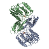
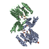
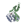
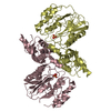
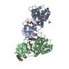
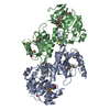
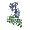
| Title | Structure of a novel N-acyl-enzyme intermediate of an N-terminal nucleophile (Ntn) hydrolase, OAT2 | ||||||
 Components Components |
| ||||||
 Keywords Keywords |  TRANSFERASE / CYTOPLASM / ACYL ENZYME / NTN HYDROLASE / ACYLTRANSFERASE / ORNITHINE ACETYL TRANSFERASE TRANSFERASE / CYTOPLASM / ACYL ENZYME / NTN HYDROLASE / ACYLTRANSFERASE / ORNITHINE ACETYL TRANSFERASE | ||||||
| Function / homology |  Function and homology informationglutamate N-acetyltransferase / glutamate N-acetyltransferase activity / acetyl-CoA:L-glutamate N-acetyltransferase activity / methione N-acyltransferase activity / amino-acid N-acetyltransferase / clavulanic acid biosynthetic process / arginine biosynthetic process / cytoplasm Function and homology informationglutamate N-acetyltransferase / glutamate N-acetyltransferase activity / acetyl-CoA:L-glutamate N-acetyltransferase activity / methione N-acyltransferase activity / amino-acid N-acetyltransferase / clavulanic acid biosynthetic process / arginine biosynthetic process / cytoplasmSimilarity search - Function | ||||||
| Biological species |  STREPTOMYCES CLAVULIGERUS (bacteria) STREPTOMYCES CLAVULIGERUS (bacteria) | ||||||
| Method | X-RAY DIFFRACTION / SYNCHROTRON / MOLECULAR REPLACEMENT / Resolution: 2.2 Å | ||||||
 Authors Authors | Iqbal, A. / Clifton, I.J. / Schofield, C.J. | ||||||
 Citation Citation | Journal: To be Published Title: Structure of a Novel N-Acyl-Enzyme Intermediate of an N-Terminal Nucleophile (Ntn) Hydrolase, Oat2 Authors: Iqbal, A. / Schofield, C.J. / Clifton, I.J. #1: Journal: Biochem.J. / Year: 2005Title: X-Ray Crystal Structure of Ornithine Acetyltransferase from the Clavulanic Acid Biosynthesis Gene Cluster. Authors: Elkins, J.M. / Kershaw, N.J. / Schofield, C.J. | ||||||
| History |
|
- Structure visualization
| Structure viewer | Molecule: MolmilJmol/JSmol |
|---|
- Downloads & links
-Download
| PDBx/mmCIF format | 2v4i.cif.gz | 282.8 KB | Display | PDBx/mmCIF format |
|---|---|---|---|---|
| PDB format | pdb2v4i.ent.gz | 226.7 KB | Display | PDB format |
| PDBx/mmJSON format | 2v4i.json.gz | Tree view | PDBx/mmJSON format | |
| Others |  Other downloads Other downloads |
-Validation report
| Arichive directory | https://data.pdbj.org/pub/pdb/validation_reports/v4/2v4iftp://data.pdbj.org/pub/pdb/validation_reports/v4/2v4i | HTTPS FTP |
|---|
-Related structure data
| Related structure data | 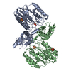 1vz6S S: Starting model for refinement |
|---|---|
| Similar structure data |


-Links
 PDBj
PDBj- Assembly
| Deposited unit | 
| |||||||||||||||||||||||||||||||||||||||||||||||||||||||||||||||||||||||||||||||||||||
|---|---|---|---|---|---|---|---|---|---|---|---|---|---|---|---|---|---|---|---|---|---|---|---|---|---|---|---|---|---|---|---|---|---|---|---|---|---|---|---|---|---|---|---|---|---|---|---|---|---|---|---|---|---|---|---|---|---|---|---|---|---|---|---|---|---|---|---|---|---|---|---|---|---|---|---|---|---|---|---|---|---|---|---|---|---|---|
| 1 | 
| |||||||||||||||||||||||||||||||||||||||||||||||||||||||||||||||||||||||||||||||||||||
| 2 | 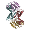
| |||||||||||||||||||||||||||||||||||||||||||||||||||||||||||||||||||||||||||||||||||||
| Unit cell |
| |||||||||||||||||||||||||||||||||||||||||||||||||||||||||||||||||||||||||||||||||||||
| Noncrystallographic symmetry (NCS) | NCS domain:
NCS domain segments:
NCS ensembles :
NCS oper:
|
-Components
| #1: Protein | Mass: 18089.531 Da / Num. of mol.: 4 / Fragment: RESIDUES 8-180 Source method: isolated from a genetically manipulated source Source: (gene. exp.) STREPTOMYCES CLAVULIGERUS (bacteria) / Plasmid: PTYB12, PET24A / Production host: ESCHERICHIA COLI (E. coli) / Strain (production host): BL21(DE3)References: UniProt: Q53940, UniProt: P0DJQ5*PLUS, glutamate N-acetyltransferase#2: Protein | Mass: 22846.332 Da / Num. of mol.: 4 / Mutation: YES Source method: isolated from a genetically manipulated source Details: ACETYLATION OF TERMINAL AMINE OF ALA 181 IN ALL FOUR CHAINS Source: (gene. exp.) STREPTOMYCES CLAVULIGERUS (bacteria) / Plasmid: PTYB12, PET24A / Production host: ESCHERICHIA COLI (E. coli) / Strain (production host): BL21(DE3)References: UniProt: Q53940, UniProt: P0DJQ5*PLUS, glutamate N-acetyltransferase#3: Water | ChemComp-HOH / | Water Mass: 18.015 Da / Num. of mol.: 301 / Source method: isolated from a natural source / Formula: H2O Mass: 18.015 Da / Num. of mol.: 301 / Source method: isolated from a natural source / Formula: H2OCompound details | ENGINEERED RESIDUE IN CHAIN B, THR 181 TO ALA ENGINEERED RESIDUE IN CHAIN D, THR 181 TO ALA ...ENGINEERED | Sequence details | T181A MUTATION | |
|---|
-Experimental details
-Experiment
| Experiment | Method: X-RAY DIFFRACTION / Number of used crystals: 1 |
|---|
- Sample preparation
| Crystal | Density Matthews: 2.61 Å3/Da / Density % sol: 52.54 % / Description: NONE |
|---|---|
| Crystal grow | pH: 7.5 Details: 1.4 M AMMONIUM SULPHATE, 0.025 M NACL, 0.1M HEPES-NA PH 7.5, 0.1 M NAG, 20 MM CDCL2 |
-Data collection
| Diffraction | Mean temperature: 100 K |
|---|---|
| Diffraction source | Source: SYNCHROTRON / Site: Diamond  / Beamline: I04 / Wavelength: 0.9699 / Beamline: I04 / Wavelength: 0.9699 |
| Detector | Type: ADSC CCD / Detector: CCD / Date: Nov 14, 2007 / Details: MIRRORS |
| Radiation | Monochromator: DOUBLE CRYSTAL / Protocol: SINGLE WAVELENGTH / Monochromatic (M) / Laue (L): M / Scattering type: x-ray |
| Radiation wavelength | Wavelength: 0.9699 Å / Relative weight: 1 |
| Reflection twin | Operator: h,-k,-l / Fraction: 0.471 |
| Reflection | Resolution: 2.2→29.7 Å / Num. obs: 79849 / % possible obs: 99.6 % / Redundancy: 3.6 % / Biso Wilson estimate: 33.4 Å2 / Rmerge(I) obs: 0.26 / Net I/σ(I): 6 |
| Reflection shell | Resolution: 2.2→2.32 Å / Redundancy: 3.6 % / Rmerge(I) obs: 0.9 / Mean I/σ(I) obs: 1.4 / % possible all: 100 |
- Processing
| Software |
| |||||||||||||||||||||||||||||||||||||||||||||||||||||||||||||||||||||||||||||||||||||||||||||||||||||||||||||||||||||||||||||||||||||||||||||||||||
|---|---|---|---|---|---|---|---|---|---|---|---|---|---|---|---|---|---|---|---|---|---|---|---|---|---|---|---|---|---|---|---|---|---|---|---|---|---|---|---|---|---|---|---|---|---|---|---|---|---|---|---|---|---|---|---|---|---|---|---|---|---|---|---|---|---|---|---|---|---|---|---|---|---|---|---|---|---|---|---|---|---|---|---|---|---|---|---|---|---|---|---|---|---|---|---|---|---|---|---|---|---|---|---|---|---|---|---|---|---|---|---|---|---|---|---|---|---|---|---|---|---|---|---|---|---|---|---|---|---|---|---|---|---|---|---|---|---|---|---|---|---|---|---|---|---|---|---|---|
| Refinement | Method to determine structure: MOLECULAR REPLACEMENT Starting model: PDB ENTRY 1VZ6 Resolution: 2.2→29.7 Å / σ(F): 1.36 / Phase error: 23.9 / Stereochemistry target values: TWIN_LSQ_F Details: TWINNING INFORMATION FRACTION 0.471 OPERATOR H,-K,-L
| |||||||||||||||||||||||||||||||||||||||||||||||||||||||||||||||||||||||||||||||||||||||||||||||||||||||||||||||||||||||||||||||||||||||||||||||||||
| Solvent computation | Shrinkage radii: 0.9 Å / VDW probe radii: 1.11 Å / Solvent model: FLAT BULK SOLVENT MODEL / Bsol: 52.984 Å2 / ksol: 0.343 e/Å3 | |||||||||||||||||||||||||||||||||||||||||||||||||||||||||||||||||||||||||||||||||||||||||||||||||||||||||||||||||||||||||||||||||||||||||||||||||||
| Displacement parameters | Biso mean: 33 Å2
| |||||||||||||||||||||||||||||||||||||||||||||||||||||||||||||||||||||||||||||||||||||||||||||||||||||||||||||||||||||||||||||||||||||||||||||||||||
| Refinement step | Cycle: LAST / Resolution: 2.2→29.7 Å
| |||||||||||||||||||||||||||||||||||||||||||||||||||||||||||||||||||||||||||||||||||||||||||||||||||||||||||||||||||||||||||||||||||||||||||||||||||
| Refine LS restraints |
| |||||||||||||||||||||||||||||||||||||||||||||||||||||||||||||||||||||||||||||||||||||||||||||||||||||||||||||||||||||||||||||||||||||||||||||||||||
| Refine LS restraints NCS |
| |||||||||||||||||||||||||||||||||||||||||||||||||||||||||||||||||||||||||||||||||||||||||||||||||||||||||||||||||||||||||||||||||||||||||||||||||||
| LS refinement shell |
|
