 Movie
Movie Controller

[English] 日本語
 Yorodumi
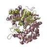
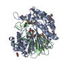
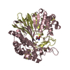
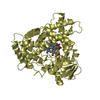
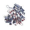
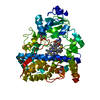
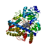
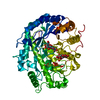
- PDB-2v36: Crystal structure of gamma-glutamyl transferase from Bacillus subtilis -
+ Open data

- Basic information
| Entry | Database: PDB / ID: 2v36 | ||||||
|---|---|---|---|---|---|---|---|
| Title | Crystal structure of gamma-glutamyl transferase from Bacillus subtilis | ||||||
 Components Components |
| ||||||
 Keywords Keywords |  TRANSFERASE / GLUTATHIONE BIOSYNTHESIS / GAMMA-GLUTAMYL TRANSFERASE / GAMMA-GLUTAMYL TRANSPEPTIDASE / ACYLTRANSFERASE / ZYMOGEN TRANSFERASE / GLUTATHIONE BIOSYNTHESIS / GAMMA-GLUTAMYL TRANSFERASE / GAMMA-GLUTAMYL TRANSPEPTIDASE / ACYLTRANSFERASE / ZYMOGEN | ||||||
| Function / homology |  Function and homology informationgamma-glutamyltransferase / glutathione gamma-glutamate hydrolase / glutathione hydrolase activity / leukotriene C4 gamma-glutamyl transferase activity / glutathione catabolic process / glutathione biosynthetic process / proteolysis / extracellular region Function and homology informationgamma-glutamyltransferase / glutathione gamma-glutamate hydrolase / glutathione hydrolase activity / leukotriene C4 gamma-glutamyl transferase activity / glutathione catabolic process / glutathione biosynthetic process / proteolysis / extracellular regionSimilarity search - Function | ||||||
| Biological species |  Bacillus subtilis (bacteria) Bacillus subtilis (bacteria) | ||||||
| Method | X-RAY DIFFRACTION / SYNCHROTRON / MOLECULAR REPLACEMENT / Resolution: 1.85 Å | ||||||
 Authors Authors | Sharath, B. / Prabhune, A.A. / Suresh, C.G. / Wilkinson, A.J. / Brannigan, J.A. | ||||||
 Citation Citation | Journal: To be Published Title: Crystal Structure of Gamma-Glutamyl Transferase Authors: Sharath, B. / Prabhune, A.A. / Suresh, C.G. / Wilkinson, A.J. / Brannigan, J.A. | ||||||
| History |
|
- Structure visualization
| Structure viewer | Molecule: MolmilJmol/JSmol |
|---|
- Downloads & links
-Download
| PDBx/mmCIF format | 2v36.cif.gz | 216.1 KB | Display | PDBx/mmCIF format |
|---|---|---|---|---|
| PDB format | pdb2v36.ent.gz | 170.7 KB | Display | PDB format |
| PDBx/mmJSON format | 2v36.json.gz | Tree view | PDBx/mmJSON format | |
| Others |  Other downloads Other downloads |
-Validation report
| Arichive directory | https://data.pdbj.org/pub/pdb/validation_reports/v3/2v36ftp://data.pdbj.org/pub/pdb/validation_reports/v3/2v36 | HTTPS FTP |
|---|
-Related structure data
| Related structure data | 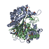 2dbuS S: Starting model for refinement |
|---|---|
| Similar structure data |

-Links
 PDBj
PDBj


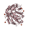
- Assembly
| Deposited unit | 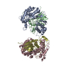
| ||||||||
|---|---|---|---|---|---|---|---|---|---|
| 1 | 
| ||||||||
| 2 | 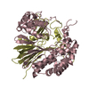
| ||||||||
| Unit cell |
|
-Components
| #1: Protein | Mass: 41452.953 Da / Num. of mol.: 2 / Fragment: RESIDUES 29-402 Source method: isolated from a genetically manipulated source Source: (gene. exp.) Bacillus subtilis (bacteria) / Plasmid: PET26B / Production host: Escherichia coli BL21(DE3) / References: UniProt: P54422, gamma-glutamyltransferase#2: Protein | Mass: 21100.697 Da / Num. of mol.: 2 Source method: isolated from a genetically manipulated source Source: (gene. exp.) Bacillus subtilis (bacteria) / Plasmid: PET26B / Production host: Escherichia coli BL21(DE3) / References: UniProt: P54422, gamma-glutamyltransferase#3: Water | ChemComp-HOH / | Water Mass: 18.015 Da / Num. of mol.: 349 / Source method: isolated from a natural source / Formula: H2O Mass: 18.015 Da / Num. of mol.: 349 / Source method: isolated from a natural source / Formula: H2O |
|---|
-Experimental details
-Experiment
| Experiment | Method: X-RAY DIFFRACTION / Number of used crystals: 1 |
|---|
- Sample preparation
| Crystal | Density Matthews: 2.7 Å3/Da / Density % sol: 54.2 % / Description: NONE |
|---|---|
| Crystal grow | pH: 7.5 Details: 0.1M SODIUM HEPES PH 7.5, 25% PEG 2000K MME AND 0.2M KSCN |
-Data collection
| Diffraction | Mean temperature: 100 K |
|---|---|
| Diffraction source | Source: SYNCHROTRON / Site: ESRF  / Beamline: ID14-1 / Wavelength: 1 / Beamline: ID14-1 / Wavelength: 1 |
| Detector | Type: ADSC CCD / Detector: CCD |
| Radiation | Protocol: SINGLE WAVELENGTH / Monochromatic (M) / Laue (L): M / Scattering type: x-ray |
| Radiation wavelength | Wavelength: 1 Å / Relative weight: 1 |
| Reflection | Resolution: 1.84→29.6 Å / Num. obs: 1952022 / % possible obs: 97.8 % / Observed criterion σ(I): 4.5 / Redundancy: 4.4 % / Rmerge(I) obs: 0.1 / Net I/σ(I): 13.8 |
| Reflection shell | Resolution: 1.85→1.92 Å / Redundancy: 2.6 % / Rmerge(I) obs: 0.44 / Mean I/σ(I) obs: 1.9 / % possible all: 82.7 |
- Processing
| Software |
| ||||||||||||||||||||
|---|---|---|---|---|---|---|---|---|---|---|---|---|---|---|---|---|---|---|---|---|---|
| Refinement | Method to determine structure: MOLECULAR REPLACEMENT Starting model: PDB ENTRY 2DBU Resolution: 1.85→90.17 Å / Cor.coef. Fo:Fc: 0.923 / Cor.coef. Fo:Fc free: 0.905 / SU B: 2.916 / SU ML: 0.091 / Cross valid method: THROUGHOUT / ESU R: 0.119 / ESU R Free: 0.135 / Stereochemistry target values: MAXIMUM LIKELIHOOD Details: HYDROGENS HAVE BEEN ADDED IN THE RIDING POSITIONS. DISORDERED REGIONS NOT MODELED
| ||||||||||||||||||||
| Solvent computation | Ion probe radii: 0.8 Å / Shrinkage radii: 0.8 Å / VDW probe radii: 1.4 Å / Solvent model: MASK | ||||||||||||||||||||
| Displacement parameters | Biso mean: 17.48 Å2
| ||||||||||||||||||||
| Refinement step | Cycle: LAST / Resolution: 1.85→90.17 Å
| ||||||||||||||||||||
| LS refinement shell | Resolution: 1.84→1.89 Å / Total num. of bins used: 20 /
|
