 Movie
Movie Controller


+ Open data

- Basic information
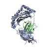
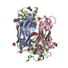
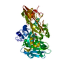
| Entry | Database: PDB / ID: 2rkk | ||||||
|---|---|---|---|---|---|---|---|
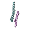
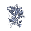
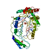
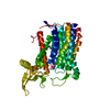
| Title | Crystal Structure of S.cerevisiae Vta1 N-terminal domain | ||||||
 Components Components | Vacuolar protein sorting-associated protein VTA1 Vacuole Vacuole | ||||||
 Keywords Keywords | LIPID TRANSPORT / MIT motif / Cytoplasm / Endosome / Membrane / Protein transport / Transport | ||||||
| Function / homology |  Function and homology informationESCRT IV complex / Endosomal Sorting Complex Required For Transport (ESCRT) / late endosome to vacuole transport via multivesicular body sorting pathway / late endosome to vacuole transport / lipid transport / ATPase activator activity / multivesicular body / protein transport / protein-macromolecule adaptor activity / endosome Function and homology informationESCRT IV complex / Endosomal Sorting Complex Required For Transport (ESCRT) / late endosome to vacuole transport via multivesicular body sorting pathway / late endosome to vacuole transport / lipid transport / ATPase activator activity / multivesicular body / protein transport / protein-macromolecule adaptor activity / endosomeSimilarity search - Function | ||||||
| Biological species |  Saccharomyces cerevisiae (brewer's yeast) Saccharomyces cerevisiae (brewer's yeast) | ||||||
| Method | X-RAY DIFFRACTION / SYNCHROTRON / SIRAS / Resolution: 2.9 Å | ||||||
 Authors Authors | Xiao, J. / Xia, H. / Zhou, J. / Xu, Z. | ||||||
 Citation Citation | Journal: Dev.Cell / Year: 2008 Title: Structural basis of vta1 function in the multivesicular body sorting pathway. Authors: Xiao, J. / Xia, H. / Zhou, J. / Azmi, I.F. / Davies, B.A. / Katzmann, D.J. / Xu, Z. | ||||||
| History |
|
- Structure visualization
| Structure viewer | Molecule: MolmilJmol/JSmol |
|---|
- Downloads & links
-Download
| PDBx/mmCIF format | 2rkk.cif.gz | 72.4 KB | Display | PDBx/mmCIF format |
|---|---|---|---|---|
| PDB format | pdb2rkk.ent.gz | 55 KB | Display | PDB format |
| PDBx/mmJSON format | 2rkk.json.gz | Tree view | PDBx/mmJSON format | |
| Others |  Other downloads Other downloads |
-Validation report
| Arichive directory | https://data.pdbj.org/pub/pdb/validation_reports/rk/2rkkftp://data.pdbj.org/pub/pdb/validation_reports/rk/2rkk | HTTPS FTP |
|---|
-Related structure data


-Links
 PDBj
PDBj
- Assembly
| Deposited unit | 
| ||||||||
|---|---|---|---|---|---|---|---|---|---|
| 1 |
| ||||||||
| Unit cell |
|
-Components
| #1: Protein | Vacuole / VPS20-associated protein 1 Mass: 19149.123 Da / Num. of mol.: 2 / Fragment: N-terminal domain Source method: isolated from a genetically manipulated source Source: (gene. exp.) Saccharomyces cerevisiae (brewer's yeast)Gene: VTA1 / Species (production host): Escherichia coli / Production host:  Escherichia coli BL21(DE3) (bacteria) / Strain (production host): BL21(DE3) / References: UniProt: Q06263 Escherichia coli BL21(DE3) (bacteria) / Strain (production host): BL21(DE3) / References: UniProt: Q06263#2: Water | ChemComp-HOH / | Water Mass: 18.015 Da / Num. of mol.: 11 / Source method: isolated from a natural source / Formula: H2O Mass: 18.015 Da / Num. of mol.: 11 / Source method: isolated from a natural source / Formula: H2O |
|---|
-Experimental details
-Experiment
| Experiment | Method: X-RAY DIFFRACTION / Number of used crystals: 1 |
|---|
- Sample preparation
| Crystal | Density Matthews: 3.65 Å3/Da / Density % sol: 66.33 % |
|---|---|
| Crystal grow | Temperature: 293 K / Method: vapor diffusion / pH: 7 Details: Sodium Formate, vapor diffusion, temperature 293K, pH 7, VAPOR DIFFUSION |
-Data collection
| Diffraction | Mean temperature: 100 K | |||||||||||||||||||||||||||||||||||||||||||||||||||||||||||||||||||||||||||||
|---|---|---|---|---|---|---|---|---|---|---|---|---|---|---|---|---|---|---|---|---|---|---|---|---|---|---|---|---|---|---|---|---|---|---|---|---|---|---|---|---|---|---|---|---|---|---|---|---|---|---|---|---|---|---|---|---|---|---|---|---|---|---|---|---|---|---|---|---|---|---|---|---|---|---|---|---|---|---|
| Diffraction source | Source: SYNCHROTRON / Site: APS  / Beamline: 21-ID-D / Wavelength: 1 Å / Beamline: 21-ID-D / Wavelength: 1 Å | |||||||||||||||||||||||||||||||||||||||||||||||||||||||||||||||||||||||||||||
| Detector | Detector: CCD / Date: Feb 8, 2007 | |||||||||||||||||||||||||||||||||||||||||||||||||||||||||||||||||||||||||||||
| Radiation | Protocol: SINGLE WAVELENGTH / Monochromatic (M) / Laue (L): M / Scattering type: x-ray | |||||||||||||||||||||||||||||||||||||||||||||||||||||||||||||||||||||||||||||
| Radiation wavelength | Wavelength: 1 Å / Relative weight: 1 | |||||||||||||||||||||||||||||||||||||||||||||||||||||||||||||||||||||||||||||
| Reflection | Redundancy: 9.7 % / Av σ(I) over netI: 11 / Number: 101963 / Rmerge(I) obs: 0.081 / Χ2: 2.13 / D res high: 3 Å / D res low: 50 Å / Num. obs: 10527 / % possible obs: 95.2 | |||||||||||||||||||||||||||||||||||||||||||||||||||||||||||||||||||||||||||||
| Diffraction reflection shell |
| |||||||||||||||||||||||||||||||||||||||||||||||||||||||||||||||||||||||||||||
| Reflection | Resolution: 2.9→50 Å / Num. obs: 12169 / % possible obs: 99.5 % / Redundancy: 7.4 % / Rmerge(I) obs: 0.062 / Χ2: 1.325 / Net I/σ(I): 11.7 | |||||||||||||||||||||||||||||||||||||||||||||||||||||||||||||||||||||||||||||
| Reflection shell |
|
-Phasing
| Phasing | Method: SIRAS |
|---|
- Processing
| Software |
| ||||||||||||||||||||||||||||
|---|---|---|---|---|---|---|---|---|---|---|---|---|---|---|---|---|---|---|---|---|---|---|---|---|---|---|---|---|---|
| Refinement | Method to determine structure: SIRAS / Resolution: 2.9→50 Å / FOM work R set: 0.79 / σ(F): 0
| ||||||||||||||||||||||||||||
| Solvent computation | Bsol: 64.107 Å2 | ||||||||||||||||||||||||||||
| Displacement parameters | Biso mean: 81.482 Å2
| ||||||||||||||||||||||||||||
| Refinement step | Cycle: LAST / Resolution: 2.9→50 Å
| ||||||||||||||||||||||||||||
| Refine LS restraints |
| ||||||||||||||||||||||||||||
| Xplor file |
|
