 Movie
Movie Controller


+ Open data

- Basic information
| Entry | Database: PDB / ID: 2rkc | ||||||
|---|---|---|---|---|---|---|---|
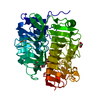
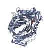
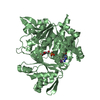
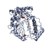
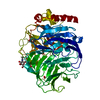
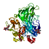
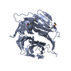
| Title | Crystal structure of the measles virus hemagglutinin | ||||||
 Components Components | Hemagglutinin | ||||||
 Keywords Keywords | VIRAL PROTEIN / hemagglutinin / measles virus / Envelope protein / Membrane / Transmembrane / Virion | ||||||
| Function / homology |  Function and homology information Function and homology informationmembrane => GO:0016020 / host cell surface receptor binding / symbiont entry into host cell / viral envelope / virion attachment to host cell / host cell plasma membrane / virion membrane / membraneSimilarity search - Function | ||||||
| Biological species |   Measles virus Measles virus | ||||||
| Method | X-RAY DIFFRACTION / SYNCHROTRON / SIRAS / Resolution: 2.7 Å | ||||||
 Authors Authors | Colf, L.A. / Juo, Z.S. / Garcia, K.C. | ||||||
 Citation Citation | Journal: To be Published Title: Structure of the measles virus hemagglutinin: implications for host cell receptor attachment. Authors: Colf, L.A. / Juo, Z.S. / Garcia, K.C. | ||||||
| History |
|
- Structure visualization
| Structure viewer | Molecule: MolmilJmol/JSmol |
|---|
- Downloads & links
-Download
| PDBx/mmCIF format | 2rkc.cif.gz | 89.5 KB | Display | PDBx/mmCIF format |
|---|---|---|---|---|
| PDB format | pdb2rkc.ent.gz | 71.1 KB | Display | PDB format |
| PDBx/mmJSON format | 2rkc.json.gz | Tree view | PDBx/mmJSON format | |
| Others |  Other downloads Other downloads |
-Validation report
| Arichive directory | https://data.pdbj.org/pub/pdb/validation_reports/rk/2rkcftp://data.pdbj.org/pub/pdb/validation_reports/rk/2rkc | HTTPS FTP |
|---|
-Related structure data
| Similar structure data |
|---|


-Links
 PDBj
PDBj


- Assembly
| Deposited unit | 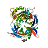
| ||||||||
|---|---|---|---|---|---|---|---|---|---|
| 1 |
| ||||||||
| Unit cell |
|
-Components
| #1: Protein | Mass: 51232.430 Da / Num. of mol.: 1 / Fragment: Extracellular domain (156-617) / Mutation: N238I Source method: isolated from a genetically manipulated source Source: (gene. exp.) Measles virus / Genus: Morbillivirus / Strain: Edmonston strain / Gene: Hemagglutinin / Plasmid: pVL1393 / Production host:   Spodoptera frugiperda (fall armyworm) / Strain (production host): Hi 5 / References: UniProt: Q83531, UniProt: P08362*PLUS Spodoptera frugiperda (fall armyworm) / Strain (production host): Hi 5 / References: UniProt: Q83531, UniProt: P08362*PLUS | ||
|---|---|---|---|
| #2: Sugar | N-Acetylglucosamine  Type: D-saccharide, beta linking / Mass: 221.208 Da / Num. of mol.: 2 Type: D-saccharide, beta linking / Mass: 221.208 Da / Num. of mol.: 2Source method: isolated from a genetically manipulated source Formula: C8H15NO6 #3: Water | ChemComp-HOH / | Water Mass: 18.015 Da / Num. of mol.: 66 / Source method: isolated from a natural source / Formula: H2O Mass: 18.015 Da / Num. of mol.: 66 / Source method: isolated from a natural source / Formula: H2O |
-Experimental details
-Experiment
| Experiment | Method: X-RAY DIFFRACTION / Number of used crystals: 2 |
|---|
- Sample preparation
| Crystal | Density Matthews: 4.42 Å3/Da / Density % sol: 72.19 % |
|---|---|
| Crystal grow | Temperature: 295 K / Method: vapor diffusion, sitting drop Details: 1.2M sodium dihydrogen phosphate, 0.3M di-potassium hydrogen phosphate, VAPOR DIFFUSION, SITTING DROP, temperature 295K |
-Data collection
| Diffraction | Mean temperature: 100 K |
|---|---|
| Diffraction source | Source: SYNCHROTRON / Site: ALS  / Beamline: 8.2.2 / Wavelength: 1.072, 1.072 / Beamline: 8.2.2 / Wavelength: 1.072, 1.072 |
| Detector | Type: ADSC QUANTUM 315 / Detector: CCD / Date: Sep 11, 2007 |
| Radiation | Monochromator: Si 111 channel / Protocol: SINGLE WAVELENGTH / Monochromatic (M) / Laue (L): M / Scattering type: x-ray |
| Radiation wavelength | Wavelength: 1.072 Å / Relative weight: 1 |
| Reflection | Resolution: 2.7→40 Å / Num. all: 48443 / Num. obs: 25665 / % possible obs: 98.8 % / Observed criterion σ(F): 2 / Observed criterion σ(I): 2 / Redundancy: 4.6 % / Biso Wilson estimate: 58.7 Å2 / Rfree details: 0.05 / Rmerge(I) obs: 0.083 / Net I/σ(I): 15 |
| Reflection shell | Resolution: 2.7→2.85 Å / Redundancy: 4.6 % / Rmerge(I) obs: 0.538 / Mean I/σ(I) obs: 2.9 / Num. unique all: 7174 / Rsym value: 0.538 / % possible all: 99.9 |
- Processing
| Software |
| |||||||||||||||||||||||||
|---|---|---|---|---|---|---|---|---|---|---|---|---|---|---|---|---|---|---|---|---|---|---|---|---|---|---|
| Refinement | Method to determine structure: SIRAS / Resolution: 2.7→40 Å / Cross valid method: THROUGHOUT / σ(F): 0 / σ(I): 0 / Stereochemistry target values: Engh & Huber
| |||||||||||||||||||||||||
| Refinement step | Cycle: LAST / Resolution: 2.7→40 Å
| |||||||||||||||||||||||||
| Refine LS restraints |
|
