 Movie
Movie Controller

[English] 日本語
 Yorodumi
- PDB-2hzh: Crystal structure of laccase from Coriolus zonatus at 2.6 A resolution -
+ Open data

- Basic information
| Entry | Database: PDB / ID: 2hzh | |||||||||
|---|---|---|---|---|---|---|---|---|---|---|
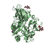
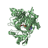
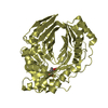
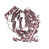
| Title | Crystal structure of laccase from Coriolus zonatus at 2.6 A resolution | |||||||||
 Components Components | laccase | |||||||||
 Keywords Keywords | OXIDOREDUCTASE / blue multi-copper enzyme / laccase from Coriolus zonatus / purification / crystals / X-ray analyses | |||||||||
| Function / homology |  Function and homology information Function and homology informationhydroquinone:oxygen oxidoreductase activity / laccase / copper ion binding / extracellular regionSimilarity search - Function | |||||||||
| Biological species |  Trametes ochracea (fungus) Trametes ochracea (fungus) | |||||||||
| Method | X-RAY DIFFRACTION / SYNCHROTRON / MOLECULAR REPLACEMENT / Resolution: 2.6 Å | |||||||||
 Authors Authors | Lyashenko, A.V. / Mikhailov, A.M. | |||||||||
 Citation Citation | Journal: To be published Title: Crystal structure of laccase from Coriolus zonatus at 2.6 A resolution Authors: Lyashenko, A.V. / Zhukhlistova, N.E. / Gabdoulkhakov, A.G. / Zhukova, Y.N. / Voelter, W. / Zaitsev, V.N. / Bento, I. / Stepanova, E.V. / Kachalova, G.S. / Koroleva, O.V. / Betzel, C. / ...Authors: Lyashenko, A.V. / Zhukhlistova, N.E. / Gabdoulkhakov, A.G. / Zhukova, Y.N. / Voelter, W. / Zaitsev, V.N. / Bento, I. / Stepanova, E.V. / Kachalova, G.S. / Koroleva, O.V. / Betzel, C. / Lindley, P.F. / Mikhailov, A.M. / Tishkov, V.I. / Morgunova, E.Y. | |||||||||
| History |
| |||||||||
| Remark 999 | SEQUENCE A UNP REFERENCE SEQUENCE FOR THE PROTEIN WAS NOT AVAILABLE AT THE TIME OF PROCESSING. |
- Structure visualization
| Structure viewer | Molecule: MolmilJmol/JSmol |
|---|
- Downloads & links
-Download
| PDBx/mmCIF format | 2hzh.cif.gz | 104.8 KB | Display | PDBx/mmCIF format |
|---|---|---|---|---|
| PDB format | pdb2hzh.ent.gz | 83.7 KB | Display | PDB format |
| PDBx/mmJSON format | 2hzh.json.gz | Tree view | PDBx/mmJSON format | |
| Others |  Other downloads Other downloads |
-Validation report
| Arichive directory | https://data.pdbj.org/pub/pdb/validation_reports/hz/2hzhftp://data.pdbj.org/pub/pdb/validation_reports/hz/2hzh | HTTPS FTP |
|---|
-Related structure data
| Similar structure data |
|---|

-Links
 PDBj
PDBj


- Assembly
| Deposited unit | 
| ||||||||
|---|---|---|---|---|---|---|---|---|---|
| 1 |
| ||||||||
| Unit cell |
| ||||||||
| Details | The biological assembly is a monomer in the asymmetric unit |
-Components
-Protein , 1 types, 1 molecules A
| #1: Protein | Mass: 52802.980 Da / Num. of mol.: 1 / Source method: isolated from a natural source / Source: (natural) Trametes ochracea (fungus) / References: UniProt: Q8TG94*PLUS |
|---|
-Sugars , 3 types, 5 molecules 


| #2: Sugar | N-Acetylglucosamine Type: D-saccharide, alpha linking / Mass: 221.208 Da / Num. of mol.: 2 Type: D-saccharide, alpha linking / Mass: 221.208 Da / Num. of mol.: 2Source method: isolated from a genetically manipulated source Formula: C8H15NO6 #3: Sugar | ChemComp-MAN / | Mannose Type: D-saccharide, alpha linking / Mass: 180.156 Da / Num. of mol.: 1 Type: D-saccharide, alpha linking / Mass: 180.156 Da / Num. of mol.: 1Source method: isolated from a genetically manipulated source Formula: C6H12O6 #4: Sugar | N-Acetylglucosamine Type: D-saccharide, beta linking / Mass: 221.208 Da / Num. of mol.: 2 Type: D-saccharide, beta linking / Mass: 221.208 Da / Num. of mol.: 2Source method: isolated from a genetically manipulated source Formula: C8H15NO6 |
|---|
-Non-polymers , 2 types, 120 molecules 

| #5: Chemical | ChemComp-CU / Copper Mass: 63.546 Da / Num. of mol.: 4 / Source method: obtained synthetically / Formula: Cu Mass: 63.546 Da / Num. of mol.: 4 / Source method: obtained synthetically / Formula: Cu#6: Water | ChemComp-HOH / | WaterMass: 18.015 Da / Num. of mol.: 116 / Source method: isolated from a natural source / Formula: H2O |
|---|
-Experimental details
-Experiment
| Experiment | Method: X-RAY DIFFRACTION / Number of used crystals: 1 |
|---|
- Sample preparation
| Crystal | Density Matthews: 5.36 Å3/Da / Density % sol: 76.86 % |
|---|---|
| Crystal grow | Temperature: 277 K / Method: vapor diffusion, hanging drop / pH: 5.5 Details: The crystallizing solution (volume 6 ml) contained the protein at a concentration of 8 mg/ml in 50 mM sodium citrate, 0.1 M ammonium sulfate, and 12.5% (w/v) PEG 4000 in 0.05M sodium acetate ...Details: The crystallizing solution (volume 6 ml) contained the protein at a concentration of 8 mg/ml in 50 mM sodium citrate, 0.1 M ammonium sulfate, and 12.5% (w/v) PEG 4000 in 0.05M sodium acetate buffer at pH 4.6., pH 5.5, VAPOR DIFFUSION, HANGING DROP, temperature 277K |
-Data collection
| Diffraction | Mean temperature: 100 K |
|---|---|
| Diffraction source | Source: SYNCHROTRON / Site: EMBL/DESY, HAMBURG  / Beamline: BW7B / Wavelength: 1.05 Å / Beamline: BW7B / Wavelength: 1.05 Å |
| Detector | Type: MAR CCD 165 mm / Detector: CCD / Date: Apr 6, 2006 |
| Radiation | Monochromator: GRAPHITE / Protocol: SINGLE WAVELENGTH / Monochromatic (M) / Laue (L): M / Scattering type: x-ray |
| Radiation wavelength | Wavelength: 1.05 Å / Relative weight: 1 |
| Reflection | Resolution: 2.6→145.86 Å / Num. obs: 35011 / % possible obs: 0.9507 % / Observed criterion σ(F): 0 / Observed criterion σ(I): 0 / Redundancy: 5.02 % |
| Reflection shell | Resolution: 2.6→2.62 Å / % possible all: 95.2 |
- Processing
| Software |
| |||||||||||||||||||||||||||||||||||||||||||||||||||||||||||||||||||||||||||||||||||||||||||||||
|---|---|---|---|---|---|---|---|---|---|---|---|---|---|---|---|---|---|---|---|---|---|---|---|---|---|---|---|---|---|---|---|---|---|---|---|---|---|---|---|---|---|---|---|---|---|---|---|---|---|---|---|---|---|---|---|---|---|---|---|---|---|---|---|---|---|---|---|---|---|---|---|---|---|---|---|---|---|---|---|---|---|---|---|---|---|---|---|---|---|---|---|---|---|---|---|---|
| Refinement | Method to determine structure: MOLECULAR REPLACEMENT / Resolution: 2.6→145.86 Å / Cor.coef. Fo:Fc: 0.914 / Cor.coef. Fo:Fc free: 0.895 / SU B: 7.652 / SU ML: 0.164 / Cross valid method: THROUGHOUT / σ(F): 0 / ESU R: 0.281 / ESU R Free: 0.225 / Stereochemistry target values: MAXIMUM LIKELIHOOD / Details: HYDROGENS HAVE BEEN ADDED IN THE RIDING POSITIONS
| |||||||||||||||||||||||||||||||||||||||||||||||||||||||||||||||||||||||||||||||||||||||||||||||
| Solvent computation | Ion probe radii: 0.8 Å / Shrinkage radii: 0.8 Å / VDW probe radii: 1.4 Å / Solvent model: MASK | |||||||||||||||||||||||||||||||||||||||||||||||||||||||||||||||||||||||||||||||||||||||||||||||
| Displacement parameters | Biso mean: 28.797 Å2
| |||||||||||||||||||||||||||||||||||||||||||||||||||||||||||||||||||||||||||||||||||||||||||||||
| Refinement step | Cycle: LAST / Resolution: 2.6→145.86 Å
| |||||||||||||||||||||||||||||||||||||||||||||||||||||||||||||||||||||||||||||||||||||||||||||||
| Refine LS restraints |
| |||||||||||||||||||||||||||||||||||||||||||||||||||||||||||||||||||||||||||||||||||||||||||||||
| LS refinement shell | Resolution: 2.6→2.668 Å / Total num. of bins used: 20
|
