SEQUENCE THE CONSTRUCT WAS EXPRESSED WITH A PURIFICATION TAG MGSDKIHHHHHHENLYFQG. THE TAG WAS ... SEQUENCE THE CONSTRUCT WAS EXPRESSED WITH A PURIFICATION TAG MGSDKIHHHHHHENLYFQG. THE TAG WAS REMOVED WITH TEV PROTEASE LEAVING ONLY A GLYCINE FOLLOWED BY THE TARGET SEQUENCE.
Mass: 18.015 Da / Num. of mol.: 202 / Source method: isolated from a natural source / Formula: H2O
Sequence details
REMARK 999 REMARK 999 SEQUENCE: THE CONSTRUCT WAS EXPRESSED WITH A PURIFICATION REMARK 999 TAG ...REMARK 999 REMARK 999 SEQUENCE: THE CONSTRUCT WAS EXPRESSED WITH A PURIFICATION REMARK 999 TAG MGSDKIHHHHHHENLYFQG. THE TAG WAS REMOVED WITH TEV PROTEASE REMARK 999 LEAVING ONLY A GLYCINE FOLLOWED BY THE TARGET SEQUENCE.
-
Experimental details
-
Experiment
Experiment
Method: X-RAY DIFFRACTION / Number of used crystals: 1
-
Sample preparation
Crystal
Density Matthews: 6.01 Å3/Da / Density % sol: 79.53 %
Crystal grow
Temperature: 277 K / Method: vapor diffusion, sitting drop / pH: 5 Details: NANODROP, 1.6M (NH4)2SO4, 0.1M Citrate pH 5.0, VAPOR DIFFUSION, SITTING DROP, temperature 277K
Resolution: 2.1→29.185 Å / Cor.coef. Fo:Fc: 0.966 / Cor.coef. Fo:Fc free: 0.954 / SU B: 5.432 / SU ML: 0.074 / TLS residual ADP flag: LIKELY RESIDUAL / Cross valid method: THROUGHOUT / σ(F): 0 / ESU R: 0.101 / ESU R Free: 0.105 / Stereochemistry target values: MAXIMUM LIKELIHOOD Details: 1. HYDROGENS HAVE BEEN ADDED IN THE RIDING POSITIONS. 2. ATOM RECORD CONTAINS RESIDUAL B FACTORS ONLY. 3. A MET-INHIBITION PROTOCOL WAS USED FOR SELENOMETHIONINE INCORPORATION DURING PROTEIN ...Details: 1. HYDROGENS HAVE BEEN ADDED IN THE RIDING POSITIONS. 2. ATOM RECORD CONTAINS RESIDUAL B FACTORS ONLY. 3. A MET-INHIBITION PROTOCOL WAS USED FOR SELENOMETHIONINE INCORPORATION DURING PROTEIN EXPRESSION. THE OCCUPANCY OF THE SE ATOMS IN THE MSE RESIDUES WAS REDUCED TO 0.75 TO ACCOUNT FOR THE REDUCED SCATTERING POWER DUE TO PARTIAL S-MET INCORPORATION. 4. SULFATE (SO4) AND CITRATE (CIT) IONS FROM THE CRYSTALLIZATION BUFFER AND GLYCEROL (GOL) FROM THE CRYO SOLUTION WERE MODELED INTO THE STRUCTURE. 5. UNEXPLAINED ELECTRON DENSITY NEAR RESIDUES 9 AND 198 WAS NOT MODELED.
Rfactor
Num. reflection
% reflection
Selection details
Rfree
0.202
1772
5 %
RANDOM
Rwork
0.17
-
-
-
obs
0.172
35315
99.65 %
-
Solvent computation
Ion probe radii: 0.8 Å / Shrinkage radii: 0.8 Å / VDW probe radii: 1.2 Å / Solvent model: MASK
Displacement parameters
Biso mean: 35.675 Å2
Baniso -1
Baniso -2
Baniso -3
1-
-1.16 Å2
0 Å2
0 Å2
2-
-
-1.16 Å2
0 Å2
3-
-
-
2.32 Å2
Refinement step
Cycle: LAST / Resolution: 2.1→29.185 Å
Protein
Nucleic acid
Ligand
Solvent
Total
Num. atoms
1612
0
88
202
1902
Refine LS restraints
Refine-ID
Type
Dev ideal
Dev ideal target
Number
X-RAY DIFFRACTION
r_bond_refined_d
0.017
0.022
1795
X-RAY DIFFRACTION
r_bond_other_d
0.002
0.02
1263
X-RAY DIFFRACTION
r_angle_refined_deg
1.649
2.01
2417
X-RAY DIFFRACTION
r_angle_other_deg
1.061
3
3052
X-RAY DIFFRACTION
r_dihedral_angle_1_deg
5.285
5
218
X-RAY DIFFRACTION
r_dihedral_angle_2_deg
28.005
22.771
83
X-RAY DIFFRACTION
r_dihedral_angle_3_deg
14.614
15
310
X-RAY DIFFRACTION
r_dihedral_angle_4_deg
22.999
15
18
X-RAY DIFFRACTION
r_chiral_restr
0.1
0.2
259
X-RAY DIFFRACTION
r_gen_planes_refined
0.006
0.02
1963
X-RAY DIFFRACTION
r_gen_planes_other
0.001
0.02
379
X-RAY DIFFRACTION
r_nbd_refined
0.212
0.2
413
X-RAY DIFFRACTION
r_nbd_other
0.197
0.2
1286
X-RAY DIFFRACTION
r_nbtor_refined
0.173
0.2
891
X-RAY DIFFRACTION
r_nbtor_other
0.085
0.2
918
X-RAY DIFFRACTION
r_xyhbond_nbd_refined
0.191
0.2
143
X-RAY DIFFRACTION
r_symmetry_vdw_refined
0.143
0.2
13
X-RAY DIFFRACTION
r_symmetry_vdw_other
0.315
0.2
41
X-RAY DIFFRACTION
r_symmetry_hbond_refined
0.243
0.2
13
X-RAY DIFFRACTION
r_mcbond_it
2.438
3
1183
X-RAY DIFFRACTION
r_mcbond_other
0.481
3
425
X-RAY DIFFRACTION
r_mcangle_it
3.212
5
1708
X-RAY DIFFRACTION
r_scbond_it
6.549
8
788
X-RAY DIFFRACTION
r_scangle_it
8.367
11
709
LS refinement shell
Resolution: 2.1→2.155 Å / Total num. of bins used: 20
Rfactor
Num. reflection
% reflection
Rfree
0.312
125
-
Rwork
0.282
2434
-
all
-
2559
-
obs
-
-
100 %
Refinement TLS params.
Method: refined / Origin x: 27.256 Å / Origin y: 11.8262 Å / Origin z: 51.5333 Å
11
12
13
21
22
23
31
32
33
T
-0.1193 Å2
0.028 Å2
0.019 Å2
-
-0.1028 Å2
-0.0263 Å2
-
-
-0.1062 Å2
L
0.8988 °2
0.0022 °2
0.0219 °2
-
1.7411 °2
0.6009 °2
-
-
1.3633 °2
S
-0.0463 Å °
0.0875 Å °
-0.1343 Å °
0.013 Å °
0.0321 Å °
-0.1521 Å °
0.1962 Å °
0.1285 Å °
0.0142 Å °
+
About Yorodumi
-
News
-
Feb 9, 2022. New format data for meta-information of EMDB entries
New format data for meta-information of EMDB entries
Version 3 of the EMDB header file is now the official format.
The previous official version 1.9 will be removed from the archive.
In the structure databanks used in Yorodumi, some data are registered as the other names, "COVID-19 virus" and "2019-nCoV". Here are the details of the virus and the list of structure data.
Jan 31, 2019. EMDB accession codes are about to change! (news from PDBe EMDB page)
EMDB accession codes are about to change! (news from PDBe EMDB page)
The allocation of 4 digits for EMDB accession codes will soon come to an end. Whilst these codes will remain in use, new EMDB accession codes will include an additional digit and will expand incrementally as the available range of codes is exhausted. The current 4-digit format prefixed with “EMD-” (i.e. EMD-XXXX) will advance to a 5-digit format (i.e. EMD-XXXXX), and so on. It is currently estimated that the 4-digit codes will be depleted around Spring 2019, at which point the 5-digit format will come into force.
The EM Navigator/Yorodumi systems omit the EMD- prefix.
Related info.:Q: What is EMD? / ID/Accession-code notation in Yorodumi/EM Navigator
Yorodumi is a browser for structure data from EMDB, PDB, SASBDB, etc.
This page is also the successor to EM Navigator detail page, and also detail information page/front-end page for Omokage search.
The word "yorodu" (or yorozu) is an old Japanese word meaning "ten thousand". "mi" (miru) is to see.
Related info.:EMDB / PDB / SASBDB / Comparison of 3 databanks / Yorodumi Search / Aug 31, 2016. New EM Navigator & Yorodumi / Yorodumi Papers / Jmol/JSmol / Function and homology information / Changes in new EM Navigator and Yorodumi
 Movie
Movie Controller

 Yorodumi
 Open data
 Basic information
 Components
 Transcriptional regulation
  Keywords
 Function and homology information
 Authors
 Citation
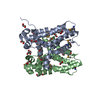
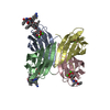
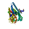
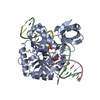
 Structure visualization
 Downloads & links
 Other downloads


 PDBj
PDBj Assembly


 Mass: 35.453 Da / Num. of mol.: 2 / Source method: obtained synthetically / Formula: Cl
 Mass: 96.063 Da / Num. of mol.: 4 / Source method: obtained synthetically / Formula: SO4
 Mass: 92.094 Da / Num. of mol.: 11 / Source method: obtained synthetically / Formula: C3H8O3
 Mass: 18.015 Da / Num. of mol.: 202 / Source method: isolated from a natural source / Formula: H2O
 Sample preparation
 / Beamline: BL11-1 / Wavelength: 1 Å
 Processing