 Movie
Movie Controller

[English] 日本語
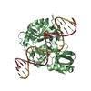
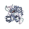
 Yorodumi
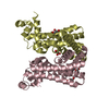
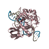
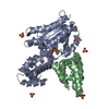
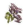
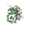
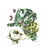
- PDB-2q2t: Structure of Chlorella virus DNA ligase-adenylate bound to a 5' p... -
+ Open data

- Basic information
| Entry | Database: PDB / ID: 2q2t | ||||||
|---|---|---|---|---|---|---|---|
| Title | Structure of Chlorella virus DNA ligase-adenylate bound to a 5' phosphorylated nick | ||||||
 Components Components |
| ||||||
 Keywords Keywords | LIGASE/DNA /  LIGASE / LYSINE ADENYLATE / PROTEIN-DNA COMPLEX / LIGASE-DNA COMPLEX LIGASE / LYSINE ADENYLATE / PROTEIN-DNA COMPLEX / LIGASE-DNA COMPLEX | ||||||
| Function / homology |  Function and homology informationDNA ligase (ATP) activity / DNA recombination / DNA replication / DNA repair / ATP binding Function and homology informationDNA ligase (ATP) activity / DNA recombination / DNA replication / DNA repair / ATP bindingSimilarity search - Function | ||||||
| Biological species |   Paramecium bursaria Chlorella virus 1 Paramecium bursaria Chlorella virus 1 | ||||||
| Method | X-RAY DIFFRACTION / SYNCHROTRON / MOLECULAR REPLACEMENT / Resolution: 2.3 Å | ||||||
 Authors Authors | Lima, C.D. / Nandakumar, J. / Nair, P.A. / Smith, P. / Shuman, S. | ||||||
 Citation Citation | Journal: Nat.Struct.Mol.Biol. / Year: 2007 Title: Structural basis for nick recognition by a minimal pluripotent DNA ligase. Authors: Nair, P.A. / Nandakumar, J. / Smith, P. / Odell, M. / Lima, C.D. / Shuman, S. | ||||||
| History |
|
- Structure visualization
| Structure viewer | Molecule: MolmilJmol/JSmol |
|---|
- Downloads & links
-Download
| PDBx/mmCIF format | 2q2t.cif.gz | 104.5 KB | Display | PDBx/mmCIF format |
|---|---|---|---|---|
| PDB format | pdb2q2t.ent.gz | 73.8 KB | Display | PDB format |
| PDBx/mmJSON format | 2q2t.json.gz | Tree view | PDBx/mmJSON format | |
| Others |  Other downloads Other downloads |
-Validation report
| Arichive directory | https://data.pdbj.org/pub/pdb/validation_reports/q2/2q2tftp://data.pdbj.org/pub/pdb/validation_reports/q2/2q2t | HTTPS FTP |
|---|
-Related structure data
| Related structure data |  2q2uC 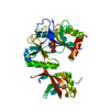 1fviS S: Starting model for refinement C: citing same article ( |
|---|---|
| Similar structure data |


-Links
 PDBj
PDBj


- Assembly
| Deposited unit | 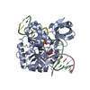
| ||||||||
|---|---|---|---|---|---|---|---|---|---|
| 1 |
| ||||||||
| Unit cell |
|
-Components
-DNA chain , 2 types, 2 molecules BD
| #1: DNA chain | Mass: 6494.194 Da / Num. of mol.: 1 / Source method: obtained synthetically / Details: Chemically synthesized. |
|---|---|
| #3: DNA chain | Mass: 3342.212 Da / Num. of mol.: 1 / Source method: obtained synthetically / Details: Chemically synthesized. |
-DNA/RNA hybrid / Protein , 2 types, 2 molecules CA
| #2: DNA/RNA hybrid | Mass: 3035.007 Da / Num. of mol.: 1 / Source method: obtained synthetically / Details: Chemically synthesized. |
|---|---|
| #4: Protein | Mass: 36833.117 Da / Num. of mol.: 1 Source method: isolated from a genetically manipulated source Source: (gene. exp.) Paramecium bursaria Chlorella virus 1 / Genus: Chlorovirus / Strain: Chlorella virus / Gene: A544R / Plasmid: pET16B / Species (production host): Escherichia coli / Production host:  Escherichia coli BL21(DE3) (bacteria) / Strain (production host): BL21(DE3) / References: UniProt: O41026 Escherichia coli BL21(DE3) (bacteria) / Strain (production host): BL21(DE3) / References: UniProt: O41026 |
-Non-polymers , 2 types, 146 molecules 

| #5: Chemical | ChemComp-AMP / Adenosine monophosphate Mass: 347.221 Da / Num. of mol.: 1 / Source method: obtained synthetically / Formula: C10H14N5O7P / Comment: AMP*YM Mass: 347.221 Da / Num. of mol.: 1 / Source method: obtained synthetically / Formula: C10H14N5O7P / Comment: AMP*YM |
|---|---|
| #6: Water | ChemComp-HOH / WaterMass: 18.015 Da / Num. of mol.: 145 / Source method: isolated from a natural source / Formula: H2O |
-Experimental details
-Experiment
| Experiment | Method: X-RAY DIFFRACTION / Number of used crystals: 1 |
|---|
- Sample preparation
| Crystal | Density Matthews: 2.59 Å3/Da / Density % sol: 52.46 % | ||||||||||||||||||||||||||||||||||||||||||||||||
|---|---|---|---|---|---|---|---|---|---|---|---|---|---|---|---|---|---|---|---|---|---|---|---|---|---|---|---|---|---|---|---|---|---|---|---|---|---|---|---|---|---|---|---|---|---|---|---|---|---|
| Crystal grow | Temperature: 295 K / Method: vapor diffusion, sitting drop / pH: 6.5 Details: A mixture of ChVLig (230 microM), nicked duplex DNA (220 microM) and 2 mM EDTA was added to an equal volume of a well solution containing 100 mM Bis-Tris-HCl (pH 6.5), 30 mM ammonium ...Details: A mixture of ChVLig (230 microM), nicked duplex DNA (220 microM) and 2 mM EDTA was added to an equal volume of a well solution containing 100 mM Bis-Tris-HCl (pH 6.5), 30 mM ammonium acetate, 22% PEG-4000. Crystals were grown at 22 C by the sitting-drop vapor diffusion method. Crystals appeared after 3 days. The crystals were transferred to buffer containing 100 mM Bis-Tris-HCl (pH 6.5), 30 mM ammonium acetate, 22% PEG-4000, 15% glycerol prior to flash-freezing in liquid nitrogen. , VAPOR DIFFUSION, SITTING DROP, temperature 295K | ||||||||||||||||||||||||||||||||||||||||||||||||
| Components of the solutions |
|
-Data collection
| Diffraction | Mean temperature: 100 K |
|---|---|
| Diffraction source | Source: SYNCHROTRON / Site: APS  / Beamline: 24-ID-C / Wavelength: 0.9792 Å / Beamline: 24-ID-C / Wavelength: 0.9792 Å |
| Detector | Type: ADSC QUANTUM 315 / Detector: CCD / Date: Mar 22, 2006 |
| Radiation | Protocol: SINGLE WAVELENGTH / Monochromatic (M) / Laue (L): M / Scattering type: x-ray |
| Radiation wavelength | Wavelength: 0.9792 Å / Relative weight: 1 |
| Reflection | Resolution: 2.3→50 Å / Num. all: 23844 / Num. obs: 23105 / % possible obs: 96.9 % / Observed criterion σ(I): -1.5 / Redundancy: 3.6 % / Rmerge(I) obs: 0.084 / Χ2: 1.05 / Net I/σ(I): 14.2 |
| Reflection shell | Resolution: 2.3→2.38 Å / Redundancy: 3.1 % / Rmerge(I) obs: 0.332 / Mean I/σ(I) obs: 3.2 / Num. unique all: 2248 / Χ2: 0.808 / % possible all: 95.7 |
- Processing
| Software |
| ||||||||||||||||||||||||||||||||||||
|---|---|---|---|---|---|---|---|---|---|---|---|---|---|---|---|---|---|---|---|---|---|---|---|---|---|---|---|---|---|---|---|---|---|---|---|---|---|
| Refinement | Method to determine structure: MOLECULAR REPLACEMENT Starting model: 1FVI Resolution: 2.3→38.99 Å / Rfactor Rfree error: 0.008 / Data cutoff high absF: 1201398.75 / Data cutoff low absF: 0 / Isotropic thermal model: RESTRAINED / Cross valid method: THROUGHOUT / σ(F): 0 / σ(I): 0 / Stereochemistry target values: Engh & Huber
| ||||||||||||||||||||||||||||||||||||
| Solvent computation | Solvent model: FLAT MODEL / Bsol: 33.819 Å2 / ksol: 0.34 e/Å3 | ||||||||||||||||||||||||||||||||||||
| Displacement parameters | Biso mean: 44.2 Å2
| ||||||||||||||||||||||||||||||||||||
| Refine analyze |
| ||||||||||||||||||||||||||||||||||||
| Refinement step | Cycle: LAST / Resolution: 2.3→38.99 Å
| ||||||||||||||||||||||||||||||||||||
| Refine LS restraints |
| ||||||||||||||||||||||||||||||||||||
| LS refinement shell | Resolution: 2.3→2.44 Å / Rfactor Rfree error: 0.03 / Total num. of bins used: 6
| ||||||||||||||||||||||||||||||||||||
| Xplor file |
|
