 Movie
Movie Controller

[English] 日本語
 Yorodumi
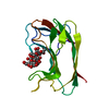
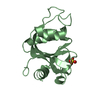
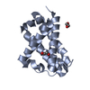
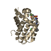
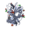
- PDB-2rg8: Crystal Structure of Programmed for Cell Death 4 Middle MA3 domain -
+ Open data

- Basic information
| Entry | Database: PDB / ID: 2rg8 | ||||||
|---|---|---|---|---|---|---|---|
| Title | Crystal Structure of Programmed for Cell Death 4 Middle MA3 domain | ||||||
 Components Components | Programmed cell death protein 4 | ||||||
 Keywords Keywords | APOPTOSIS / TRANSLATION / MA3 domain / HEAT repeats / Anti-oncogene / Cell cycle / Cytoplasm / Nucleus / Phosphorylation / Polymorphism / RNA-binding | ||||||
| Function / homology |  Function and homology information Function and homology informationepithelial to mesenchymal transition involved in cardiac fibroblast development / negative regulation of myofibroblast differentiation / negative regulation of vascular associated smooth muscle cell differentiation / negative regulation of JUN kinase activity / response to alkaloid / positive regulation of endothelial cell apoptotic process / positive regulation of vascular associated smooth muscle cell apoptotic process / negative regulation of vascular associated smooth muscle cell proliferation / negative regulation of cytokine production involved in inflammatory response / BMP signaling pathway ...epithelial to mesenchymal transition involved in cardiac fibroblast development / negative regulation of myofibroblast differentiation / negative regulation of vascular associated smooth muscle cell differentiation / negative regulation of JUN kinase activity / response to alkaloid / positive regulation of endothelial cell apoptotic process / positive regulation of vascular associated smooth muscle cell apoptotic process / negative regulation of vascular associated smooth muscle cell proliferation / negative regulation of cytokine production involved in inflammatory response / BMP signaling pathway / Gene and protein expression by JAK-STAT signaling after Interleukin-12 stimulation / response to hormone / positive regulation of inflammatory response / positive regulation of non-canonical NF-kappaB signal transduction / cellular response to lipopolysaccharide / negative regulation of DNA-templated transcription / apoptotic process / negative regulation of apoptotic process / RNA binding / nucleus / cytosol / cytoplasmSimilarity search - Function | ||||||
| Biological species |  Homo sapiens (human) Homo sapiens (human) | ||||||
| Method | X-RAY DIFFRACTION / SYNCHROTRON / SAD / Resolution: 1.8 Å | ||||||
 Authors Authors | Garces, R. / Suzuki, C. / Wagner, G. | ||||||
 Citation Citation | Journal: Proc.Natl.Acad.Sci.Usa / Year: 2008 Title: PDCD4 inhibits translation initiation by binding to eIF4A using both its MA3 domains. Authors: Suzuki, C. / Garces, R.G. / Edmonds, K.A. / Hiller, S. / Hyberts, S.G. / Marintchev, A. / Wagner, G. | ||||||
| History |
|
- Structure visualization
| Structure viewer | Molecule: MolmilJmol/JSmol |
|---|
- Downloads & links
-Download
| PDBx/mmCIF format | 2rg8.cif.gz | 75.8 KB | Display | PDBx/mmCIF format |
|---|---|---|---|---|
| PDB format | pdb2rg8.ent.gz | 60.3 KB | Display | PDB format |
| PDBx/mmJSON format | 2rg8.json.gz | Tree view | PDBx/mmJSON format | |
| Others |  Other downloads Other downloads |
-Validation report
| Arichive directory | https://data.pdbj.org/pub/pdb/validation_reports/rg/2rg8ftp://data.pdbj.org/pub/pdb/validation_reports/rg/2rg8 | HTTPS FTP |
|---|
-Related structure data
| Similar structure data |
|---|


-Links
 PDBj
PDBj


- Assembly
| Deposited unit | 
| ||||||||
|---|---|---|---|---|---|---|---|---|---|
| 1 | 
| ||||||||
| 2 | 
| ||||||||
| Unit cell |
|
-Components
| #1: Protein | / Nuclear antigen H731-like / Neoplastic transformation inhibitor protein / Protein 197/15a Mass: 18001.789 Da / Num. of mol.: 2 / Fragment: MA3 domain Source method: isolated from a genetically manipulated source Source: (gene. exp.) Homo sapiens (human) / Gene: PDCD4, H731 / Species (production host): Escherichia coli / Production host:  Escherichia coli BL21(DE3) (bacteria) / Strain (production host): BL21(DE3) / References: UniProt: Q53EL6 Escherichia coli BL21(DE3) (bacteria) / Strain (production host): BL21(DE3) / References: UniProt: Q53EL6#2: Chemical | Chloride  Mass: 35.453 Da / Num. of mol.: 2 / Source method: obtained synthetically / Formula: Cl Mass: 35.453 Da / Num. of mol.: 2 / Source method: obtained synthetically / Formula: Cl#3: Chemical |   Mass: 22.990 Da / Num. of mol.: 3 / Source method: obtained synthetically / Formula: Na Mass: 22.990 Da / Num. of mol.: 3 / Source method: obtained synthetically / Formula: Na#4: Water | ChemComp-HOH / | Water Mass: 18.015 Da / Num. of mol.: 430 / Source method: isolated from a natural source / Formula: H2O Mass: 18.015 Da / Num. of mol.: 430 / Source method: isolated from a natural source / Formula: H2O |
|---|
-Experimental details
-Experiment
| Experiment | Method: X-RAY DIFFRACTION / Number of used crystals: 2 |
|---|
- Sample preparation
| Crystal | Density Matthews: 2.21 Å3/Da / Density % sol: 44.4 % |
|---|---|
| Crystal grow | Temperature: 300 K / Method: vapor diffusion, hanging drop / pH: 8.5 Details: 200mM sodium acetate, 28%-30% PEG4000, 8% Jeffamine M-600, pH 8.5, VAPOR DIFFUSION, HANGING DROP, temperature 300K |
-Data collection
| Diffraction |
| ||||||||||||||||||
|---|---|---|---|---|---|---|---|---|---|---|---|---|---|---|---|---|---|---|---|
| Diffraction source |
| ||||||||||||||||||
| Detector |
| ||||||||||||||||||
| Radiation |
| ||||||||||||||||||
| Radiation wavelength |
| ||||||||||||||||||
| Reflection | Resolution: 1.8→35.68 Å / Num. all: 34183 / Num. obs: 34012 / % possible obs: 99.5 % / Observed criterion σ(F): 2 / Observed criterion σ(I): 2 / Redundancy: 11.7 % / Biso Wilson estimate: 14.8 Å2 / Rsym value: 0.095 |

- Processing
| Software |
| ||||||||||||||||||||||||||||||||||||||||||||||||||||||||||||||||||||||||||||||||
|---|---|---|---|---|---|---|---|---|---|---|---|---|---|---|---|---|---|---|---|---|---|---|---|---|---|---|---|---|---|---|---|---|---|---|---|---|---|---|---|---|---|---|---|---|---|---|---|---|---|---|---|---|---|---|---|---|---|---|---|---|---|---|---|---|---|---|---|---|---|---|---|---|---|---|---|---|---|---|---|---|---|
| Refinement | Method to determine structure: SAD / Resolution: 1.8→35.68 Å / Rfactor Rfree error: 0.005 / Data cutoff high absF: 85310.93 / Data cutoff low absF: 0 / Isotropic thermal model: RESTRAINED / Cross valid method: THROUGHOUT / σ(F): 0 / Stereochemistry target values: Engh & Huber / Details: BULK SOLVENT MODEL USED
| ||||||||||||||||||||||||||||||||||||||||||||||||||||||||||||||||||||||||||||||||
| Solvent computation | Solvent model: FLAT MODEL / Bsol: 41.1428 Å2 / ksol: 0.35 e/Å3 | ||||||||||||||||||||||||||||||||||||||||||||||||||||||||||||||||||||||||||||||||
| Displacement parameters | Biso mean: 18.2 Å2
| ||||||||||||||||||||||||||||||||||||||||||||||||||||||||||||||||||||||||||||||||
| Refine analyze |
| ||||||||||||||||||||||||||||||||||||||||||||||||||||||||||||||||||||||||||||||||
| Refinement step | Cycle: LAST / Resolution: 1.8→35.68 Å
| ||||||||||||||||||||||||||||||||||||||||||||||||||||||||||||||||||||||||||||||||
| Refine LS restraints |
| ||||||||||||||||||||||||||||||||||||||||||||||||||||||||||||||||||||||||||||||||
| LS refinement shell | Resolution: 1.8→1.91 Å / Rfactor Rfree error: 0.012 / Total num. of bins used: 6
| ||||||||||||||||||||||||||||||||||||||||||||||||||||||||||||||||||||||||||||||||
| Xplor file |
|
