 Movie
Movie Controller

[English] 日本語
 Yorodumi
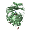
- PDB-3hfi: The crystal structure of the putative regulator from Escherichia ... -
+ Open data

- Basic information
| Entry | Database: PDB / ID: 3hfi | ||||||
|---|---|---|---|---|---|---|---|
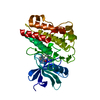
| Title | The crystal structure of the putative regulator from Escherichia coli CFT073 | ||||||
 Components Components | Putative regulator | ||||||
 Keywords Keywords |  structural genomics / unknown function / regulator / structural geonomics / PSI / MCSG / Protein Structure Initiative / Midwest Center for Structural Genomics / DNA-binding / Transcription / Transcription regulation structural genomics / unknown function / regulator / structural geonomics / PSI / MCSG / Protein Structure Initiative / Midwest Center for Structural Genomics / DNA-binding / Transcription / Transcription regulation | ||||||
| Function / homology |  Function and homology information Function and homology information | ||||||
| Biological species |  Escherichia coli O6 (bacteria) Escherichia coli O6 (bacteria) | ||||||
| Method | X-RAY DIFFRACTION / SYNCHROTRON / SAD / Resolution: 2.2 Å | ||||||
 Authors Authors | Zhang, R. / Xu, X. / Zheng, H. / Savchenko, A. / Edwards, A. / Joachimiak, A. / Midwest Center for Structural Genomics (MCSG) | ||||||
 Citation Citation | Journal: To be Published Title: The crystal structure of the putative regulator from Escherichia coli CFT073 Authors: Zhang, R. / Xu, X. / Zheng, H. / Savchenko, A. / Edwards, A. / Joachimiak, A. | ||||||
| History |
|
- Structure visualization
| Structure viewer | Molecule: MolmilJmol/JSmol |
|---|
- Downloads & links
-Download
| PDBx/mmCIF format | 3hfi.cif.gz | 41.6 KB | Display | PDBx/mmCIF format |
|---|---|---|---|---|
| PDB format | pdb3hfi.ent.gz | 29.5 KB | Display | PDB format |
| PDBx/mmJSON format | 3hfi.json.gz | Tree view | PDBx/mmJSON format | |
| Others |  Other downloads Other downloads |
-Validation report
| Arichive directory | https://data.pdbj.org/pub/pdb/validation_reports/hf/3hfiftp://data.pdbj.org/pub/pdb/validation_reports/hf/3hfi | HTTPS FTP |
|---|
-Related structure data
| Similar structure data | |
|---|---|
| Other databases |


-Links
 PDBj
PDBj- Assembly
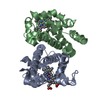
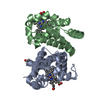
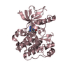
| Deposited unit | 
| ||||||||
|---|---|---|---|---|---|---|---|---|---|
| 1 | 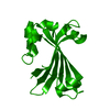
| ||||||||
| Unit cell |
| ||||||||
| Components on special symmetry positions |
| ||||||||
| Details | This protein exists as dimer. The second part of the biological assembly is generated by the axis: Y,X,-Z |
-Components
| #1: Protein | Mass: 19661.430 Da / Num. of mol.: 1 / Fragment: residues 82-251 Source method: isolated from a genetically manipulated source Source: (gene. exp.) Escherichia coli O6 (bacteria) / Strain: CFT073 / Gene: c4276, GI:26250098 / Plasmid: pMCSG7 / Production host: Escherichia coli BL21 (bacteria) / Strain (production host): BL21 / References: UniProt: Q8FCM7, UniProt: A0A0H2VBL3*PLUS |
|---|---|
| #2: Water | ChemComp-HOH / Water Mass: 18.015 Da / Num. of mol.: 40 / Source method: isolated from a natural source / Formula: H2O Mass: 18.015 Da / Num. of mol.: 40 / Source method: isolated from a natural source / Formula: H2O |
-Experimental details
-Experiment
| Experiment | Method: X-RAY DIFFRACTION / Number of used crystals: 1 |
|---|
- Sample preparation
| Crystal | Density Matthews: 2.3 Å3/Da / Density % sol: 46.42 % |
|---|---|
| Crystal grow | Temperature: 289 K / Method: vapor diffusion, sitting drop / pH: 4.6 Details: 0.1M Na(OAC), 3.5M NaFormate, 1/600 CHYMOTRYPSIN, pH 4.6, VAPOR DIFFUSION, SITTING DROP, temperature 289K |
-Data collection
| Diffraction | Mean temperature: 100 K |
|---|---|
| Diffraction source | Source: SYNCHROTRON / Site: APS  / Beamline: 19-ID / Wavelength: 0.9794 Å / Beamline: 19-ID / Wavelength: 0.9794 Å |
| Detector | Type: ADSC QUANTUM 315r / Detector: CCD / Date: Aug 8, 2008 / Details: mirrors |
| Radiation | Monochromator: Si 111 channel / Protocol: SINGLE WAVELENGTH / Monochromatic (M) / Laue (L): M / Scattering type: x-ray |
| Radiation wavelength | Wavelength: 0.9794 Å / Relative weight: 1 |
| Reflection | Resolution: 2.2→89.8 Å / Num. all: 9339 / Num. obs: 9325 / % possible obs: 99.85 % / Observed criterion σ(F): 2 / Observed criterion σ(I): 2 / Redundancy: 27.5 % / Rmerge(I) obs: 0.096 / Net I/σ(I): 51.9 |
| Reflection shell | Resolution: 2.2→2.257 Å / Redundancy: 26.6 % / Rmerge(I) obs: 0.734 / Mean I/σ(I) obs: 3.4 / % possible all: 99.3 |
- Processing
| Software |
| ||||||||||||||||||||||||||||||||||||||||||||||||||||||||||||||||||||||||||||||||||||||||||||||||||||||||||||||||||||||||||||||||||||||||||||||||||||||||||||||||||||||||||
|---|---|---|---|---|---|---|---|---|---|---|---|---|---|---|---|---|---|---|---|---|---|---|---|---|---|---|---|---|---|---|---|---|---|---|---|---|---|---|---|---|---|---|---|---|---|---|---|---|---|---|---|---|---|---|---|---|---|---|---|---|---|---|---|---|---|---|---|---|---|---|---|---|---|---|---|---|---|---|---|---|---|---|---|---|---|---|---|---|---|---|---|---|---|---|---|---|---|---|---|---|---|---|---|---|---|---|---|---|---|---|---|---|---|---|---|---|---|---|---|---|---|---|---|---|---|---|---|---|---|---|---|---|---|---|---|---|---|---|---|---|---|---|---|---|---|---|---|---|---|---|---|---|---|---|---|---|---|---|---|---|---|---|---|---|---|---|---|---|---|---|---|
| Refinement | Method to determine structure: SAD / Resolution: 2.2→89.8 Å / Cor.coef. Fo:Fc: 0.954 / Cor.coef. Fo:Fc free: 0.935 / SU B: 12.14 / SU ML: 0.14 / TLS residual ADP flag: LIKELY RESIDUAL / Cross valid method: THROUGHOUT / ESU R: 0.224 / ESU R Free: 0.202 Stereochemistry target values: MAXIMUM LIKELIHOOD WITH PHASES Details: HYDROGENS HAVE BEEN ADDED IN THE RIDING POSITIONS
| ||||||||||||||||||||||||||||||||||||||||||||||||||||||||||||||||||||||||||||||||||||||||||||||||||||||||||||||||||||||||||||||||||||||||||||||||||||||||||||||||||||||||||
| Solvent computation | Ion probe radii: 0.8 Å / Shrinkage radii: 0.8 Å / VDW probe radii: 1.2 Å / Solvent model: MASK | ||||||||||||||||||||||||||||||||||||||||||||||||||||||||||||||||||||||||||||||||||||||||||||||||||||||||||||||||||||||||||||||||||||||||||||||||||||||||||||||||||||||||||
| Displacement parameters | Biso mean: 21.47 Å2
| ||||||||||||||||||||||||||||||||||||||||||||||||||||||||||||||||||||||||||||||||||||||||||||||||||||||||||||||||||||||||||||||||||||||||||||||||||||||||||||||||||||||||||
| Refinement step | Cycle: LAST / Resolution: 2.2→89.8 Å
| ||||||||||||||||||||||||||||||||||||||||||||||||||||||||||||||||||||||||||||||||||||||||||||||||||||||||||||||||||||||||||||||||||||||||||||||||||||||||||||||||||||||||||
| Refine LS restraints |
| ||||||||||||||||||||||||||||||||||||||||||||||||||||||||||||||||||||||||||||||||||||||||||||||||||||||||||||||||||||||||||||||||||||||||||||||||||||||||||||||||||||||||||
| LS refinement shell | Resolution: 2.2→2.257 Å / Total num. of bins used: 20
| ||||||||||||||||||||||||||||||||||||||||||||||||||||||||||||||||||||||||||||||||||||||||||||||||||||||||||||||||||||||||||||||||||||||||||||||||||||||||||||||||||||||||||
| Refinement TLS params. | Method: refined / Origin x: 40.897 Å / Origin y: 46.583 Å / Origin z: 3.807 Å
| ||||||||||||||||||||||||||||||||||||||||||||||||||||||||||||||||||||||||||||||||||||||||||||||||||||||||||||||||||||||||||||||||||||||||||||||||||||||||||||||||||||||||||
| Refinement TLS group |
|
