 Movie
Movie Controller


+ Open data

- Basic information
| Entry | Database: PDB / ID: 2rf5 | ||||||
|---|---|---|---|---|---|---|---|
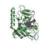
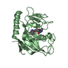
| Title | Crystal structure of human tankyrase 1- catalytic PARP domain | ||||||
 Components Components | Tankyrase-1 | ||||||
 Keywords Keywords | TRANSFERASE / CATALYTIC FRAGMENT / PARP / Structural Genomics / Structural Genomics Consortium / SGC / ADP-ribosylation / ANK repeat / Chromosomal protein / Glycosyltransferase / Golgi apparatus / Membrane / mRNA transport / NAD / Nuclear pore complex / Nucleus / Phosphorylation / Protein transport / Telomere / Translocation / Transport | ||||||
| Function / homology |  Function and homology information Function and homology informationnegative regulation of telomeric DNA binding / negative regulation of maintenance of mitotic sister chromatid cohesion, telomeric / regulation of telomere maintenance via telomerase / XAV939 stabilizes AXIN / NAD+ ADP-ribosyltransferase / negative regulation of telomere maintenance via telomere lengthening / protein auto-ADP-ribosylation / protein localization to chromosome, telomeric region / protein poly-ADP-ribosylation / pericentriolar material ...negative regulation of telomeric DNA binding / negative regulation of maintenance of mitotic sister chromatid cohesion, telomeric / regulation of telomere maintenance via telomerase / XAV939 stabilizes AXIN / NAD+ ADP-ribosyltransferase / negative regulation of telomere maintenance via telomere lengthening / protein auto-ADP-ribosylation / protein localization to chromosome, telomeric region / protein poly-ADP-ribosylation / pericentriolar material / mitotic spindle pole / NAD+-protein ADP-ribosyltransferase activity / positive regulation of telomere capping / Transferases; Glycosyltransferases; Pentosyltransferases / NAD+ ADP-ribosyltransferase activity / spindle assembly / mRNA transport / nuclear pore / positive regulation of telomerase activity / positive regulation of telomere maintenance via telomerase / nucleotidyltransferase activity / mitotic spindle organization / TCF dependent signaling in response to WNT / Degradation of AXIN / peptidyl-threonine phosphorylation / Wnt signaling pathway / Regulation of PTEN stability and activity / protein polyubiquitination / positive regulation of canonical Wnt signaling pathway / protein transport / histone binding / peptidyl-serine phosphorylation / nuclear membrane / chromosome, telomeric region / nuclear body / Ub-specific processing proteases / cell division / Golgi membrane / Golgi apparatus / positive regulation of transcription by RNA polymerase II / zinc ion binding / nucleoplasm / nucleus / cytosol / cytoplasmSimilarity search - Function | ||||||
| Biological species |  Homo sapiens (human) Homo sapiens (human) | ||||||
| Method | X-RAY DIFFRACTION / SYNCHROTRON / MOLECULAR REPLACEMENT / Resolution: 2.3 Å | ||||||
 Authors Authors | Lehtio, L. / Karlberg, T. / Arrowsmith, C.H. / Berglund, H. / Busam, R. / Collins, R. / Dahlgren, L.G. / Edwards, A.M. / Flodin, S. / Flores, A. ...Lehtio, L. / Karlberg, T. / Arrowsmith, C.H. / Berglund, H. / Busam, R. / Collins, R. / Dahlgren, L.G. / Edwards, A.M. / Flodin, S. / Flores, A. / Graslund, S. / Hammarstrom, M. / Herman, M.D. / Holmberg-Schiavone, L. / Johansson, I. / Kallas, A. / Kotenyova, T. / Moche, M. / Nordlund, P. / Nyman, T. / Persson, C. / Sagemark, J. / Sundstrom, M. / Thorsell, A.G. / Tresaugues, L. / van den Berg, S. / Welin, M. / Weigelt, J. / Structural Genomics Consortium (SGC) | ||||||
 Citation Citation | Journal: J.Mol.Biol. / Year: 2008 Title: Zinc binding catalytic domain of human tankyrase 1. Authors: Lehtio, L. / Collins, R. / van den Berg, S. / Johansson, A. / Dahlgren, L.G. / Hammarstrom, M. / Helleday, T. / Holmberg-Schiavone, L. / Karlberg, T. / Weigelt, J. | ||||||
| History |
|
- Structure visualization
| Structure viewer | Molecule: MolmilJmol/JSmol |
|---|
- Downloads & links
-Download
| PDBx/mmCIF format | 2rf5.cif.gz | 59.9 KB | Display | PDBx/mmCIF format |
|---|---|---|---|---|
| PDB format | pdb2rf5.ent.gz | 41.7 KB | Display | PDB format |
| PDBx/mmJSON format | 2rf5.json.gz | Tree view | PDBx/mmJSON format | |
| Others |  Other downloads Other downloads |
-Validation report
| Arichive directory | https://data.pdbj.org/pub/pdb/validation_reports/rf/2rf5ftp://data.pdbj.org/pub/pdb/validation_reports/rf/2rf5 | HTTPS FTP |
|---|
-Related structure data
| Related structure data |  2pqfS S: Starting model for refinement |
|---|---|
| Similar structure data |


-Links
 PDBj
PDBj


- Assembly
| Deposited unit | 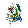
| ||||||||
|---|---|---|---|---|---|---|---|---|---|
| 1 |
| ||||||||
| Unit cell |
| ||||||||
| Details | Authors state that the asymmetric unit is a biological monomer of this domain. |
-Components
| #1: Protein | / TANK1 / Tankyrase I / TNKS-1 / TRF1- interacting ankyrin-related ADP-ribose polymerase Mass: 29417.082 Da / Num. of mol.: 1 / Fragment: PARP catalytic domain: Residues 1091-1325 / Mutation: M1266I Source method: isolated from a genetically manipulated source Source: (gene. exp.) Homo sapiens (human) / Gene: TNKS, PARP5A, PARPL, TIN1, TINF1, TNKS1 / Plasmid: pNIC-Bsa4 / Production host:  Escherichia coli (E. coli) / Strain (production host): BL21(DE3)Rosetta2 / References: UniProt: O95271, NAD+ ADP-ribosyltransferase Escherichia coli (E. coli) / Strain (production host): BL21(DE3)Rosetta2 / References: UniProt: O95271, NAD+ ADP-ribosyltransferase |
|---|---|
| #2: Chemical | ChemComp-ZN /   Mass: 65.409 Da / Num. of mol.: 1 / Source method: obtained synthetically / Formula: Zn Mass: 65.409 Da / Num. of mol.: 1 / Source method: obtained synthetically / Formula: Zn |
| #3: Chemical | ChemComp-GOL / Glycerol  Mass: 92.094 Da / Num. of mol.: 1 / Source method: obtained synthetically / Formula: C3H8O3 Mass: 92.094 Da / Num. of mol.: 1 / Source method: obtained synthetically / Formula: C3H8O3 |
| #4: Water | ChemComp-HOH / Water Mass: 18.015 Da / Num. of mol.: 96 / Source method: isolated from a natural source / Formula: H2O Mass: 18.015 Da / Num. of mol.: 96 / Source method: isolated from a natural source / Formula: H2O |
-Experimental details
-Experiment
| Experiment | Method: X-RAY DIFFRACTION / Number of used crystals: 2 |
|---|
- Sample preparation
| Crystal | Density Matthews: 2.28 Å3/Da / Density % sol: 46.12 % |
|---|---|
| Crystal grow | Temperature: 293 K / Method: vapor diffusion, sitting drop / pH: 5.9 Details: 18% PEG 3350, 0.18M Mg formate, 3% Hexanediol, pH 5.9, VAPOR DIFFUSION, SITTING DROP, temperature 293K |
-Data collection
| Diffraction | Mean temperature: 100 K |
|---|---|
| Diffraction source | Source: SYNCHROTRON / Site: BESSY  / Beamline: 14.1 / Wavelength: 0.97981 Å / Beamline: 14.1 / Wavelength: 0.97981 Å |
| Detector | Type: MARMOSAIC 225 mm CCD / Detector: CCD / Date: May 16, 2007 / Details: mirrors |
| Radiation | Monochromator: Si(111) / Protocol: SINGLE WAVELENGTH / Monochromatic (M) / Laue (L): M / Scattering type: x-ray |
| Radiation wavelength | Wavelength: 0.97981 Å / Relative weight: 1 |
| Reflection | Resolution: 2.3→20 Å / Num. all: 12043 / Num. obs: 12043 / % possible obs: 97.8 % / Observed criterion σ(F): 0 / Observed criterion σ(I): 0 / Redundancy: 9 % / Biso Wilson estimate: 30.6 Å2 / Rmerge(I) obs: 0.166 / Net I/σ(I): 13.39 |
| Reflection shell | Resolution: 2.3→2.4 Å / Redundancy: 7.5 % / Rmerge(I) obs: 0.47 / Mean I/σ(I) obs: 4.21 / Num. unique all: 1448 / % possible all: 99.9 |
- Processing
| Software |
| ||||||||||||||||||||||||||||||||||||||||||||||||||||||||||||||||||||||||||||||||||||||||||||||||||||||||||||||||||||||||||||||||||||||||||||||||||||||||||||||||||||||||||
|---|---|---|---|---|---|---|---|---|---|---|---|---|---|---|---|---|---|---|---|---|---|---|---|---|---|---|---|---|---|---|---|---|---|---|---|---|---|---|---|---|---|---|---|---|---|---|---|---|---|---|---|---|---|---|---|---|---|---|---|---|---|---|---|---|---|---|---|---|---|---|---|---|---|---|---|---|---|---|---|---|---|---|---|---|---|---|---|---|---|---|---|---|---|---|---|---|---|---|---|---|---|---|---|---|---|---|---|---|---|---|---|---|---|---|---|---|---|---|---|---|---|---|---|---|---|---|---|---|---|---|---|---|---|---|---|---|---|---|---|---|---|---|---|---|---|---|---|---|---|---|---|---|---|---|---|---|---|---|---|---|---|---|---|---|---|---|---|---|---|---|---|
| Refinement | Method to determine structure: MOLECULAR REPLACEMENT Starting model: PDB entry 2PQF Resolution: 2.3→19.44 Å / Cor.coef. Fo:Fc: 0.932 / Cor.coef. Fo:Fc free: 0.871 / SU B: 12.238 / SU ML: 0.159 / TLS residual ADP flag: LIKELY RESIDUAL / Isotropic thermal model: ISOTROPIC AND TLS / Cross valid method: THROUGHOUT / σ(F): 0 / ESU R: 0.304 / ESU R Free: 0.238 / Stereochemistry target values: MAXIMUM LIKELIHOOD
| ||||||||||||||||||||||||||||||||||||||||||||||||||||||||||||||||||||||||||||||||||||||||||||||||||||||||||||||||||||||||||||||||||||||||||||||||||||||||||||||||||||||||||
| Solvent computation | Ion probe radii: 0.8 Å / Shrinkage radii: 0.8 Å / VDW probe radii: 1.2 Å / Solvent model: MASK | ||||||||||||||||||||||||||||||||||||||||||||||||||||||||||||||||||||||||||||||||||||||||||||||||||||||||||||||||||||||||||||||||||||||||||||||||||||||||||||||||||||||||||
| Displacement parameters | Biso mean: 22.62 Å2
| ||||||||||||||||||||||||||||||||||||||||||||||||||||||||||||||||||||||||||||||||||||||||||||||||||||||||||||||||||||||||||||||||||||||||||||||||||||||||||||||||||||||||||
| Refinement step | Cycle: LAST / Resolution: 2.3→19.44 Å
| ||||||||||||||||||||||||||||||||||||||||||||||||||||||||||||||||||||||||||||||||||||||||||||||||||||||||||||||||||||||||||||||||||||||||||||||||||||||||||||||||||||||||||
| Refine LS restraints |
| ||||||||||||||||||||||||||||||||||||||||||||||||||||||||||||||||||||||||||||||||||||||||||||||||||||||||||||||||||||||||||||||||||||||||||||||||||||||||||||||||||||||||||
| LS refinement shell | Resolution: 2.3→2.359 Å / Total num. of bins used: 20
| ||||||||||||||||||||||||||||||||||||||||||||||||||||||||||||||||||||||||||||||||||||||||||||||||||||||||||||||||||||||||||||||||||||||||||||||||||||||||||||||||||||||||||
| Refinement TLS params. | Method: refined / Refine-ID: X-RAY DIFFRACTION
| ||||||||||||||||||||||||||||||||||||||||||||||||||||||||||||||||||||||||||||||||||||||||||||||||||||||||||||||||||||||||||||||||||||||||||||||||||||||||||||||||||||||||||
| Refinement TLS group |
|
