 Movie
Movie Controller

[English] 日本語
 Yorodumi
- PDB-2rd9: Crystal structure of a putative yfit-like metal-dependent hydrola... -
+ Open data

- Basic information
| Entry | Database: PDB / ID: 2rd9 | ||||||
|---|---|---|---|---|---|---|---|
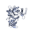
| Title | Crystal structure of a putative yfit-like metal-dependent hydrolase (bh0186) from bacillus halodurans c-125 at 2.30 A resolution | ||||||
 Components Components | BH0186 protein | ||||||
 Keywords Keywords |  HYDROLASE / Structural genomics / Joint Center for Structural Genomics / JCSG / Protein Structure Initiative / PSI-2 HYDROLASE / Structural genomics / Joint Center for Structural Genomics / JCSG / Protein Structure Initiative / PSI-2 | ||||||
| Function / homology |  Function and homology information Function and homology informationDinB-like domain / DinB superfamily / dinb family like domain / DinB/YfiT-like putative metalloenzymes / Four Helix Bundle (Hemerythrin (Met), subunit A) / Up-down Bundle / Mainly Alpha Similarity search - Domain/homology | ||||||
| Biological species |  Bacillus halodurans C-125 (bacteria) Bacillus halodurans C-125 (bacteria) | ||||||
| Method | X-RAY DIFFRACTION / SYNCHROTRON / MAD / Resolution: 2.3 Å | ||||||
 Authors Authors | Joint Center for Structural Genomics (JCSG) | ||||||
 Citation Citation | Journal: To be published Title: Crystal structure of YfiT-like putative metal-dependent hydrolase (NP_241052.1) from Bacillus halodurans at 2.30 A resolution Authors: Joint Center for Structural Genomics (JCSG) | ||||||
| History |
| ||||||
| Remark 300 | BIOMOLECULE: 1, 2, 3 SEE REMARK 350 FOR THE AUTHOR PROVIDED AND PROGRAM GENERATED ASSEMBLY ... BIOMOLECULE: 1, 2, 3 SEE REMARK 350 FOR THE AUTHOR PROVIDED AND PROGRAM GENERATED ASSEMBLY INFORMATION FOR THE STRUCTURE IN THIS ENTRY. THE REMARK MAY ALSO PROVIDE INFORMATION ON BURIED SURFACE AREA. SIZE EXCLUSION CHROMATOGRAPHY SUPPORTS THE ASSIGNMENT OF A DIMER AS THE SIGNIFICANT OLIGOMERIZATION STATE. | ||||||
| Remark 999 | SEQUENCE THE CONSTRUCT WAS EXPRESSED WITH A PURIFICATION TAG MGSDKIHHHHHHENLYFQG. |
- Structure visualization
| Structure viewer | Molecule: MolmilJmol/JSmol |
|---|
- Downloads & links
-Download
| PDBx/mmCIF format | 2rd9.cif.gz | 182.1 KB | Display | PDBx/mmCIF format |
|---|---|---|---|---|
| PDB format | pdb2rd9.ent.gz | 147.7 KB | Display | PDB format |
| PDBx/mmJSON format | 2rd9.json.gz | Tree view | PDBx/mmJSON format | |
| Others |  Other downloads Other downloads |
-Validation report
| Arichive directory | https://data.pdbj.org/pub/pdb/validation_reports/rd/2rd9ftp://data.pdbj.org/pub/pdb/validation_reports/rd/2rd9 | HTTPS FTP |
|---|
-Related structure data
| Similar structure data | |
|---|---|
| Other databases |


-Links
 PDBj
PDBj


- Assembly
| Deposited unit | 
| ||||||||||||||||||||||||||||||||||||||||||||||||||||||||||||||||||||||||||||||||||||||||||||||||||||||
|---|---|---|---|---|---|---|---|---|---|---|---|---|---|---|---|---|---|---|---|---|---|---|---|---|---|---|---|---|---|---|---|---|---|---|---|---|---|---|---|---|---|---|---|---|---|---|---|---|---|---|---|---|---|---|---|---|---|---|---|---|---|---|---|---|---|---|---|---|---|---|---|---|---|---|---|---|---|---|---|---|---|---|---|---|---|---|---|---|---|---|---|---|---|---|---|---|---|---|---|---|---|---|---|
| 1 | 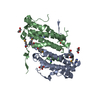
| ||||||||||||||||||||||||||||||||||||||||||||||||||||||||||||||||||||||||||||||||||||||||||||||||||||||
| 2 | 
| ||||||||||||||||||||||||||||||||||||||||||||||||||||||||||||||||||||||||||||||||||||||||||||||||||||||
| 3 |
| ||||||||||||||||||||||||||||||||||||||||||||||||||||||||||||||||||||||||||||||||||||||||||||||||||||||
| Unit cell |
| ||||||||||||||||||||||||||||||||||||||||||||||||||||||||||||||||||||||||||||||||||||||||||||||||||||||
| Noncrystallographic symmetry (NCS) | NCS domain:
NCS domain segments: Component-ID: 1 / Refine code: 4
NCS ensembles :
| ||||||||||||||||||||||||||||||||||||||||||||||||||||||||||||||||||||||||||||||||||||||||||||||||||||||
| Details | SIZE EXCLUSION CHROMATOGRAPHY SUPPORTS THE ASSIGNMENT OF A DIMER AS THE SIGNIFICANT OLIGOMERIZATION STATE. |
-Components
| #1: Protein | Mass: 22890.209 Da / Num. of mol.: 4 Source method: isolated from a genetically manipulated source Source: (gene. exp.) Bacillus halodurans C-125 (bacteria) / Species: Bacillus halodurans / Strain: C-125, DSM 18197, FERM 7344, JCM 9153 / Gene: NP_241052.1, BH0186 / Plasmid: speedET / Production host: Escherichia coli (E. coli) / Strain (production host): HK100 / References: UniProt: Q9KGB8#2: Chemical | ChemComp-NI / Nickel  Mass: 58.693 Da / Num. of mol.: 4 / Source method: obtained synthetically / Formula: Ni Mass: 58.693 Da / Num. of mol.: 4 / Source method: obtained synthetically / Formula: Ni#3: Chemical | ChemComp-IMD / Imidazole  Mass: 69.085 Da / Num. of mol.: 5 / Source method: obtained synthetically / Formula: C3H5N2 Mass: 69.085 Da / Num. of mol.: 5 / Source method: obtained synthetically / Formula: C3H5N2#4: Chemical | ChemComp-GOL / Glycerol  Mass: 92.094 Da / Num. of mol.: 7 / Source method: obtained synthetically / Formula: C3H8O3 Mass: 92.094 Da / Num. of mol.: 7 / Source method: obtained synthetically / Formula: C3H8O3#5: Water | ChemComp-HOH / | Water Mass: 18.015 Da / Num. of mol.: 555 / Source method: isolated from a natural source / Formula: H2O Mass: 18.015 Da / Num. of mol.: 555 / Source method: isolated from a natural source / Formula: H2O |
|---|
-Experimental details
-Experiment
| Experiment | Method: X-RAY DIFFRACTION / Number of used crystals: 1 |
|---|
- Sample preparation
| Crystal | Density Matthews: 2.91 Å3/Da / Density % sol: 57.74 % |
|---|---|
| Crystal grow | Temperature: 277 K / Method: vapor diffusion, sitting drop / pH: 7 Details: NANODROP, 10.0% PEG 6000, 0.1M HEPES pH 7.0, VAPOR DIFFUSION, SITTING DROP, temperature 277K |
-Data collection
| Diffraction | Mean temperature: 100 K | |||||||||||||||||||||||||||||||||||||||||||||||||||||||||||||||||||||||||||||
|---|---|---|---|---|---|---|---|---|---|---|---|---|---|---|---|---|---|---|---|---|---|---|---|---|---|---|---|---|---|---|---|---|---|---|---|---|---|---|---|---|---|---|---|---|---|---|---|---|---|---|---|---|---|---|---|---|---|---|---|---|---|---|---|---|---|---|---|---|---|---|---|---|---|---|---|---|---|---|
| Diffraction source | Source: SYNCHROTRON / Site: SSRL  / Beamline: BL11-1 / Wavelength: 0.91837, 0.97941 / Beamline: BL11-1 / Wavelength: 0.91837, 0.97941 | |||||||||||||||||||||||||||||||||||||||||||||||||||||||||||||||||||||||||||||
| Detector | Type: MARMOSAIC 325 mm CCD / Detector: CCD / Date: Jul 20, 2007 / Details: Flat mirror (vertical focusing) | |||||||||||||||||||||||||||||||||||||||||||||||||||||||||||||||||||||||||||||
| Radiation | Monochromator: Single crystal Si(111) bent (horizontal focusing) Protocol: MAD / Monochromatic (M) / Laue (L): M / Scattering type: x-ray | |||||||||||||||||||||||||||||||||||||||||||||||||||||||||||||||||||||||||||||
| Radiation wavelength |
| |||||||||||||||||||||||||||||||||||||||||||||||||||||||||||||||||||||||||||||
| Reflection | Resolution: 2.3→29.463 Å / Num. obs: 47316 / % possible obs: 99.5 % / Observed criterion σ(I): -3 / Biso Wilson estimate: 36.076 Å2 / Rmerge(I) obs: 0.066 / Net I/σ(I): 14.51 | |||||||||||||||||||||||||||||||||||||||||||||||||||||||||||||||||||||||||||||
| Reflection shell |
|
-Phasing
| Phasing | Method: MAD |
|---|
- Processing
| Software |
| |||||||||||||||||||||||||||||||||||||||||||||||||||||||||||||||||||||||||||||||||||||||||||||||||||||||||||||||||||||||||||||
|---|---|---|---|---|---|---|---|---|---|---|---|---|---|---|---|---|---|---|---|---|---|---|---|---|---|---|---|---|---|---|---|---|---|---|---|---|---|---|---|---|---|---|---|---|---|---|---|---|---|---|---|---|---|---|---|---|---|---|---|---|---|---|---|---|---|---|---|---|---|---|---|---|---|---|---|---|---|---|---|---|---|---|---|---|---|---|---|---|---|---|---|---|---|---|---|---|---|---|---|---|---|---|---|---|---|---|---|---|---|---|---|---|---|---|---|---|---|---|---|---|---|---|---|---|---|---|
| Refinement | Method to determine structure: MAD / Resolution: 2.3→29.463 Å / Cor.coef. Fo:Fc: 0.961 / Cor.coef. Fo:Fc free: 0.936 / SU B: 9.357 / SU ML: 0.126 / TLS residual ADP flag: LIKELY RESIDUAL / Cross valid method: THROUGHOUT / σ(F): 0 / ESU R: 0.227 / ESU R Free: 0.185 Stereochemistry target values: MAXIMUM LIKELIHOOD WITH PHASES Details: 1. HYDROGENS HAVE BEEN ADDED IN THE RIDING POSITIONS. 2. ATOM RECORDS CONTAIN RESIDUAL B FACTORS ONLY. 3. A MET-INHIBITION PROTOCOL WAS USED FOR SELENOMETHIONINE INCORPORATION DURING PROTEIN ...Details: 1. HYDROGENS HAVE BEEN ADDED IN THE RIDING POSITIONS. 2. ATOM RECORDS CONTAIN RESIDUAL B FACTORS ONLY. 3. A MET-INHIBITION PROTOCOL WAS USED FOR SELENOMETHIONINE INCORPORATION DURING PROTEIN EXPRESSION. THE OCCUPANCY OF THE SE ATOMS IN THE MSE RESIDUES WAS REDUCED TO 0.75 FOR THE REDUCED SCATTERING POWER DUE TO PARTIAL S-MET INCORPORATION. 4. RESIDUES 77-84 IN CHAIN B ARE DISORDERED AND NOT INCLUDED IN THE MODEL. 5. A NICKEL ION IS COORDINATED IN AN OCTAHEDRAL COMPLEX TO THE SIDECHAINS OF HIS48, HIS142 AND HIS146 ON THE SAME CHAIN, HIS-9 ON THE NEIGHBORING CHAIN, AND ONE IMIDAZOLE AND ONE WATER MOLECULES. X-RAY FLUORESCENCE SUPPORTS THE ASSIGNMENT OF THE NICKEL ION. 5. IMIDAZOLE MOLECULES FROM THE PURIFICATION SOLUTION ARE MODELED. 6. GLYCEROL MOLECULES FROM THE CRYO SOLUTION ARE MODELED.
| |||||||||||||||||||||||||||||||||||||||||||||||||||||||||||||||||||||||||||||||||||||||||||||||||||||||||||||||||||||||||||||
| Solvent computation | Ion probe radii: 0.8 Å / Shrinkage radii: 0.8 Å / VDW probe radii: 1.2 Å / Solvent model: MASK | |||||||||||||||||||||||||||||||||||||||||||||||||||||||||||||||||||||||||||||||||||||||||||||||||||||||||||||||||||||||||||||
| Displacement parameters | Biso mean: 25.05 Å2 | |||||||||||||||||||||||||||||||||||||||||||||||||||||||||||||||||||||||||||||||||||||||||||||||||||||||||||||||||||||||||||||
| Refinement step | Cycle: LAST / Resolution: 2.3→29.463 Å
| |||||||||||||||||||||||||||||||||||||||||||||||||||||||||||||||||||||||||||||||||||||||||||||||||||||||||||||||||||||||||||||
| Refine LS restraints |
| |||||||||||||||||||||||||||||||||||||||||||||||||||||||||||||||||||||||||||||||||||||||||||||||||||||||||||||||||||||||||||||
| Refine LS restraints NCS | Refine-ID: X-RAY DIFFRACTION
| |||||||||||||||||||||||||||||||||||||||||||||||||||||||||||||||||||||||||||||||||||||||||||||||||||||||||||||||||||||||||||||
| LS refinement shell | Resolution: 2.3→2.36 Å / Total num. of bins used: 20
| |||||||||||||||||||||||||||||||||||||||||||||||||||||||||||||||||||||||||||||||||||||||||||||||||||||||||||||||||||||||||||||
| Refinement TLS params. | Method: refined / Refine-ID: X-RAY DIFFRACTION
| |||||||||||||||||||||||||||||||||||||||||||||||||||||||||||||||||||||||||||||||||||||||||||||||||||||||||||||||||||||||||||||
| Refinement TLS group |
|
