[English] 日本語
Yorodumi
- PDB-2ra1: Crystal structure of the N-terminal part of the bacterial S-layer... -

+
Open data


ID or keywords:

Loading...

-
Basic information

Entry
Database: PDB / ID: 2ra1
TitleCrystal structure of the N-terminal part of the bacterial S-layer protein SbsC
ComponentsSurface layer protein
KeywordsSUGAR BINDING PROTEIN / triple coiled-coil / s-layer protein / PROTEIN BINDING
Function / homology
Function and homology information


identical protein binding / metal ion binding
Similarity search - Function
Methane Monooxygenase Hydroxylase; Chain G, domain 1 - #770 / Methane Monooxygenase Hydroxylase; Chain G, domain 1 - #780 / Methane Monooxygenase Hydroxylase; Chain G, domain 1 - #790 / Immunoglobulin-like - #2120 / S-layer protein SbsC, C-terminal domain / SbsC C-terminal domain / SbsC C-terminal domain / S-layer protein SbsC C-terminal domain / Bacterial Ig-like domain / Immunoglobulin-like - #1220 ...Methane Monooxygenase Hydroxylase; Chain G, domain 1 - #770 / Methane Monooxygenase Hydroxylase; Chain G, domain 1 - #780 / Methane Monooxygenase Hydroxylase; Chain G, domain 1 - #790 / Immunoglobulin-like - #2120 / S-layer protein SbsC, C-terminal domain / SbsC C-terminal domain / SbsC C-terminal domain / S-layer protein SbsC C-terminal domain / Bacterial Ig-like domain / Immunoglobulin-like - #1220 / Copper resistance protein CopC/internalin, immunoglobulin-like / Bacterial Ig-like domain 2 / Bacterial Ig-like domain, group 2 / Methane Monooxygenase Hydroxylase; Chain G, domain 1 / Immunoglobulin-like fold / Up-down Bundle / Immunoglobulin-like / Sandwich / Mainly Beta / Mainly Alpha
Similarity search - Domain/homology
Surface layer protein
Similarity search - Component
Biological speciesGeobacillus stearothermophilus (bacteria)
MethodX-RAY DIFFRACTION / SYNCHROTRON / MOLECULAR REPLACEMENT / molecular replacement / Resolution: 2.406 Å
AuthorsPavkov, T. / Egelseer, E.M. / Tesarz, M. / Sleytr, U.B. / Keller, W.
CitationJournal: Structure / Year: 2008
Title: The structure and binding behavior of the bacterial cell surface layer protein SbsC.
Authors: Pavkov, T. / Egelseer, E.M. / Tesarz, M. / Svergun, D.I. / Sleytr, U.B. / Keller, W.
History
DepositionSep 14, 2007Deposition site: RCSB / Processing site: RCSB
Revision 1.0Aug 19, 2008Provider: repository / Type: Initial release
Revision 1.1Jul 13, 2011Group: Version format compliance
Revision 1.2Oct 25, 2017Group: Refinement description / Category: software
Revision 1.3Feb 21, 2024Group: Data collection / Database references / Category: chem_comp_atom / chem_comp_bond / database_2
Item: _database_2.pdbx_DOI / _database_2.pdbx_database_accession

-
Structure visualization

Structure viewerMolecule:
MolmilJmol/JSmol

Downloads & links

-
Assembly

Deposited unit
A: Surface layer protein


Theoretical massNumber of molelcules
Total (without water)45,0521
Polymers45,0521
Non-polymers00
Water4,540252
1


  • Idetical with deposited unit
  • defined by author&software
TypeNameSymmetry operationNumber
identity operation1_555x,y,z1
MethodPISA
Unit cell
Length a, b, c (Å)49.645, 94.877, 59.646
Angle α, β, γ (deg.)90.000, 106.680, 90.000
Int Tables number4
Space group name H-MP1211

-
Components

#1: Protein Surface layer protein /


Mass: 45051.746 Da / Num. of mol.: 1 / Fragment: N-terminal domains
Source method: isolated from a genetically manipulated source
Source: (gene. exp.) Geobacillus stearothermophilus (bacteria)
Gene: sbsC / Plasmid: pET3a / Production host: Escherichia coli (E. coli) / Strain (production host): HMS174(DE3) / References: UniProt: O68840
#2: Water ChemComp-HOH / water / Water


Mass: 18.015 Da / Num. of mol.: 252 / Source method: isolated from a natural source / Formula: H2O

-
Experimental details

-
Experiment

ExperimentMethod: X-RAY DIFFRACTION / Number of used crystals: 1

-
Sample preparation

CrystalDensity Matthews: 2.99 Å3/Da / Density % sol: 58.82 %
Crystal growTemperature: 298 K / Method: batch method / pH: 6.5
Details: 6-14% PEG3350, various salts - NaCl, LiCl etc., pH 6.5, batch method, temperature 298K
Temp details: 5.5-7.5

-
Data collection

DiffractionMean temperature: 100 K
Diffraction sourceSource: SYNCHROTRON / Site: ELETTRA / Beamline: 5.2R / Wavelength: 1.2 Å
DetectorType: MARRESEARCH / Detector: CCD / Date: Oct 15, 2005
RadiationProtocol: SINGLE WAVELENGTH / Monochromatic (M) / Laue (L): M / Scattering type: x-ray
Radiation wavelengthWavelength: 1.2 Å / Relative weight: 1
ReflectionResolution: 2.4→99 Å / Num. obs: 20541 / % possible obs: 99.7 % / Rmerge(I) obs: 0.064 / Χ2: 0.829 / Net I/σ(I): 22.1
Reflection shell
Resolution (Å)Rmerge(I) obsNum. unique allΧ2% possible all
2.4-2.440.19610051.37597.7
2.44-2.490.15510530.708100
2.49-2.530.1389890.672100
2.53-2.590.12610280.623100
2.59-2.640.13610160.906100
2.64-2.70.26810442.022100
2.7-2.770.09110010.555100
2.77-2.850.08810340.52699.9
2.85-2.930.07310370.485100
2.93-3.020.06610140.438100
3.02-3.130.0610470.41100
3.13-3.260.05410150.412100
3.26-3.410.05110150.469100
3.41-3.590.06310420.924100
3.59-3.810.06110481.015100
3.81-4.10.06810241.086100
4.1-4.520.07110201.126100
4.52-5.170.05810280.657100
5.17-6.510.05210580.72100
6.51-990.05310231.06396.1

-
Phasing

Phasing
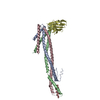
Method: molecular replacement
Phasing MRModel details: Phaser MODE: MR_AUTO
Highest resolutionLowest resolution
Rotation2.8 Å15.02 Å
Translation2.8 Å15.02 Å

-
Processing

Software
NameVersionClassificationNB
DENZOdata reduction
SCALEPACKdata scaling
PHASERphasing
PHENIXrefinement
PDB_EXTRACT3data extraction
Refinement
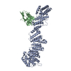
Method to determine structure: MOLECULAR REPLACEMENT
Starting model: N-terminal domains form the structure of rSbsC(31-844)

Resolution: 2.406→20.009 Å / FOM work R set: 0.811 / Stereochemistry target values: ml
RfactorNum. reflection% reflection
Rfree0.24 1036 5.16 %
Rwork0.177 --
obs-20089 97.62 %
Solvent computationBsol: 42.844 Å2 / ksol: 0.333 e/Å3
Displacement parametersBiso max: 109.03 Å2 / Biso mean: 40.03 Å2 / Biso min: 9.77 Å2
Baniso -1Baniso -2Baniso -3
1--4.18 Å2-0 Å2-3.295 Å2
2---7.269 Å20 Å2
3---11.449 Å2
Refinement stepCycle: LAST / Resolution: 2.406→20.009 Å
ProteinNucleic acidLigandSolventTotal
Num. atoms3173 0 0 252 3425
Refine LS restraints
Refine-IDTypeDev idealWeight
X-RAY DIFFRACTIONf_angle_d1.1311
X-RAY DIFFRACTIONf_bond_d0.0081
X-RAY DIFFRACTIONf_chiral_restr0.0751
X-RAY DIFFRACTIONf_dihedral_angle_d17.2181
X-RAY DIFFRACTIONf_plane_restr0.0041
X-RAY DIFFRACTIONf_nbd_refined4.1061
LS refinement shell
Resolution (Å)Rfactor RworkNum. reflection RworkRefine-IDTotal num. of bins used% reflection obs (%)
2.406-2.4280.2423X-RAY DIFFRACTION3880
2.428-2.450.216490X-RAY DIFFRACTION3889
2.45-2.4730.219463X-RAY DIFFRACTION3888
2.473-2.4970.209519X-RAY DIFFRACTION3890
2.497-2.5220.212465X-RAY DIFFRACTION3892
2.522-2.5480.194491X-RAY DIFFRACTION3890
2.548-2.5750.198487X-RAY DIFFRACTION3891
2.575-2.6030.211489X-RAY DIFFRACTION3891
2.603-2.6320.207503X-RAY DIFFRACTION3890
2.632-2.6630.188454X-RAY DIFFRACTION3889
2.663-2.6960.201504X-RAY DIFFRACTION3891
2.696-2.730.225500X-RAY DIFFRACTION3892
2.73-2.7660.208502X-RAY DIFFRACTION3892
2.766-2.8030.212508X-RAY DIFFRACTION3895
2.803-2.8430.205488X-RAY DIFFRACTION3892
2.843-2.8860.209518X-RAY DIFFRACTION3894
2.886-2.9310.206505X-RAY DIFFRACTION3895
2.931-2.9790.21504X-RAY DIFFRACTION3892
2.979-3.030.193484X-RAY DIFFRACTION3892
3.03-3.0850.211502X-RAY DIFFRACTION3895
3.085-3.1440.189530X-RAY DIFFRACTION3893
3.144-3.2080.194474X-RAY DIFFRACTION3892
3.208-3.2770.187525X-RAY DIFFRACTION3895
3.277-3.3530.181507X-RAY DIFFRACTION3896
3.353-3.4370.185495X-RAY DIFFRACTION3892
3.437-3.5290.17519X-RAY DIFFRACTION3894
3.529-3.6320.164518X-RAY DIFFRACTION3896
3.632-3.7490.159512X-RAY DIFFRACTION3896
3.749-3.8820.156510X-RAY DIFFRACTION3893
3.882-4.0360.154509X-RAY DIFFRACTION3894
4.036-4.2180.147501X-RAY DIFFRACTION3893
4.218-4.4380.133536X-RAY DIFFRACTION3896
4.438-4.7130.125498X-RAY DIFFRACTION3894
4.713-5.0710.139525X-RAY DIFFRACTION3894
5.071-5.5710.155518X-RAY DIFFRACTION3895
5.571-6.3550.171514X-RAY DIFFRACTION3895
6.355-7.9230.145523X-RAY DIFFRACTION3895
7.923-20.010.13540X-RAY DIFFRACTION3895
Refinement TLS params.

Method: refined / Refine-ID: X-RAY DIFFRACTION

IDL112)L122)L132)L222)L232)L332)S11 (Å °)S12 (Å °)S13 (Å °)S21 (Å °)S22 (Å °)S23 (Å °)S31 (Å °)S32 (Å °)S33 (Å °)T112)T122)T132)T222)T232)T332)Origin x (Å)Origin y (Å)Origin z (Å)
10.7421-0.68510.07771.36-0.25520.017-0.0714-0.07290.02560.03790.053-0.2295-0.0545-0.009600.1174-0.00990.00010.13790.03380.180376.6721-58.17751.26
20.513-0.0905-0.30340.3012-0.20230.39030.45970.44510.4934-0.1508-0.1835-0.2032-0.3534-0.25670.01350.26820.15740.02830.23740.12570.267150.9429-28.282135.4641
30.4112-0.238-0.63180.6829-0.46321.12640.1574-0.33740.17480.1635-0.114-0.16090.59560.27470.00020.37660.0569-0.04790.27960.01040.116422.94-8.788126.7294
41.22410.1503-0.84420.14270.08160.8874-0.20670.3101-0.0094-0.04620.097-0.10460.13850.00090.00010.1276-0.03250.00840.1808-0.04070.151117.87163.945658.7025
Refinement TLS group
IDRefine-IDRefine TLS-IDSelection detailsLabel asym-IDLabel seq-ID
1X-RAY DIFFRACTION1chain A and resid 32-198A32 - 198
2X-RAY DIFFRACTION2chain A and resid 199-259A199 - 259
3X-RAY DIFFRACTION3chain A and resid 262-357A262 - 357
4X-RAY DIFFRACTION4chain A and resid 360-443A360 - 443

+
About Yorodumi

-
News

-
Feb 9, 2022. New format data for meta-information of EMDB entries

New format data for meta-information of EMDB entries

  • Version 3 of the EMDB header file is now the official format.
  • The previous official version 1.9 will be removed from the archive.

Related info.:EMDB header

External links:wwPDB to switch to version 3 of the EMDB data model

-
Aug 12, 2020. Covid-19 info

Covid-19 info

URL: https://pdbj.org/emnavi/covid19.php

New page: Covid-19 featured information page in EM Navigator.

Related info.:Covid-19 info / Mar 5, 2020. Novel coronavirus structure data

+
Mar 5, 2020. Novel coronavirus structure data

Novel coronavirus structure data

Related info.:Yorodumi Speices / Aug 12, 2020. Covid-19 info

External links:COVID-19 featured content - PDBj / Molecule of the Month (242):Coronavirus Proteases

+
Jan 31, 2019. EMDB accession codes are about to change! (news from PDBe EMDB page)

EMDB accession codes are about to change! (news from PDBe EMDB page)

  • The allocation of 4 digits for EMDB accession codes will soon come to an end. Whilst these codes will remain in use, new EMDB accession codes will include an additional digit and will expand incrementally as the available range of codes is exhausted. The current 4-digit format prefixed with “EMD-” (i.e. EMD-XXXX) will advance to a 5-digit format (i.e. EMD-XXXXX), and so on. It is currently estimated that the 4-digit codes will be depleted around Spring 2019, at which point the 5-digit format will come into force.
  • The EM Navigator/Yorodumi systems omit the EMD- prefix.

Related info.:Q: What is EMD? / ID/Accession-code notation in Yorodumi/EM Navigator

External links:EMDB Accession Codes are Changing Soon! / Contact to PDBj

+
Jul 12, 2017. Major update of PDB

Major update of PDB

  • wwPDB released updated PDB data conforming to the new PDBx/mmCIF dictionary.
  • This is a major update changing the version number from 4 to 5, and with Remediation, in which all the entries are updated.
  • In this update, many items about electron microscopy experimental information are reorganized (e.g. em_software).
  • Now, EM Navigator and Yorodumi are based on the updated data.

External links:wwPDB Remediation / Enriched Model Files Conforming to OneDep Data Standards Now Available in the PDB FTP Archive

-
Yorodumi

Thousand views of thousand structures

  • Yorodumi is a browser for structure data from EMDB, PDB, SASBDB, etc.
  • This page is also the successor to EM Navigator detail page, and also detail information page/front-end page for Omokage search.
  • The word "yorodu" (or yorozu) is an old Japanese word meaning "ten thousand". "mi" (miru) is to see.

Related info.:EMDB / PDB / SASBDB / Comparison of 3 databanks / Yorodumi Search / Aug 31, 2016. New EM Navigator & Yorodumi / Yorodumi Papers / Jmol/JSmol / Function and homology information / Changes in new EM Navigator and Yorodumi

Read more