 Movie
Movie Controller

[English] 日本語
 Yorodumi
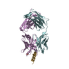
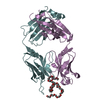
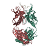
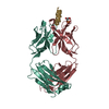
- PDB-1tzg: Crystal structure of HIV-1 neutralizing human Fab 4E10 in complex... -
+ Open data

- Basic information
| Entry | Database: PDB / ID: 1tzg | ||||||
|---|---|---|---|---|---|---|---|
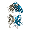
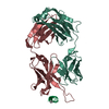
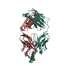
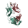
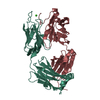
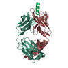
| Title | Crystal structure of HIV-1 neutralizing human Fab 4E10 in complex with a 13-residue peptide containing the 4E10 epitope on gp41 | ||||||
 Components Components |
| ||||||
 Keywords Keywords |  IMMUNE SYSTEM / antibody-epitope complex IMMUNE SYSTEM / antibody-epitope complex | ||||||
| Function / homology | Immunoglobulins / Immunoglobulin-like / Sandwich / Mainly Beta Function and homology information Function and homology information | ||||||
| Biological species |  Homo sapiens (human) Homo sapiens (human) | ||||||
| Method | X-RAY DIFFRACTION / SYNCHROTRON / MOLECULAR REPLACEMENT / Resolution: 2.2 Å | ||||||
 Authors Authors | Cardoso, R.M.F. / Zwick, M.B. / Stanfield, R.L. / Kunert, R. / Binley, J.M. / Katinger, H. / Burton, D.R. / Wilson, I.A. | ||||||
 Citation Citation | Journal: Immunity / Year: 2005 Title: Broadly Neutralizing Anti-HIV Antibody 4E10 Recognizes a Helical Conformation of a Highly Conserved Fusion-Associated Motif in gp41 Authors: Cardoso, R.M.F. / Zwick, M.B. / Stanfield, R.L. / Kunert, R. / Binley, J.M. / Katinger, H. / Burton, D.R. / Wilson, I.A. | ||||||
| History |
| ||||||
| Remark 999 | SEQUENCE A suitable database reference sequence could not be found for the protein chains in this ...SEQUENCE A suitable database reference sequence could not be found for the protein chains in this structure at the time of processing |
- Structure visualization
| Structure viewer | Molecule: MolmilJmol/JSmol |
|---|
- Downloads & links
-Download
| PDBx/mmCIF format | 1tzg.cif.gz | 197.3 KB | Display | PDBx/mmCIF format |
|---|---|---|---|---|
| PDB format | pdb1tzg.ent.gz | 155.3 KB | Display | PDB format |
| PDBx/mmJSON format | 1tzg.json.gz | Tree view | PDBx/mmJSON format | |
| Others |  Other downloads Other downloads |
-Validation report
| Arichive directory | https://data.pdbj.org/pub/pdb/validation_reports/tz/1tzgftp://data.pdbj.org/pub/pdb/validation_reports/tz/1tzg | HTTPS FTP |
|---|
-Related structure data
| Related structure data | 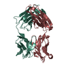 1hklS S: Starting model for refinement |
|---|---|
| Similar structure data |
-Links
 PDBj
PDBj

- Assembly
| Deposited unit | 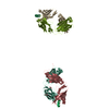
| ||||||||
|---|---|---|---|---|---|---|---|---|---|
| 1 | 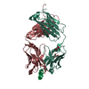
| ||||||||
| 2 | 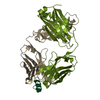
| ||||||||
| 3 | 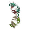
| ||||||||
| Unit cell |
| ||||||||
| Details | The asymmetric unit contains two biological units (2 Fab-peptide complexes) |
-Components
| #1: Antibody | Mass: 23292.705 Da / Num. of mol.: 2 / Fragment: light chain Source method: isolated from a genetically manipulated source Source: (gene. exp.) Homo sapiens (human) / Cell (production host): ovary / Production host:   Cricetulus griseus (Chinese hamster) Cricetulus griseus (Chinese hamster)#2: Antibody | Mass: 24424.518 Da / Num. of mol.: 2 / Fragment: heavy chain Source method: isolated from a genetically manipulated source Source: (gene. exp.) Homo sapiens (human) / Cell (production host): ovary / Production host: Cricetulus griseus (Chinese hamster)#3: Protein/peptide | Mass: 1653.837 Da / Num. of mol.: 2 / Fragment: Transmembrane glycoprotein / Source method: obtained synthetically #4: Chemical | Glycerol  Mass: 92.094 Da / Num. of mol.: 3 / Source method: obtained synthetically / Formula: C3H8O3 Mass: 92.094 Da / Num. of mol.: 3 / Source method: obtained synthetically / Formula: C3H8O3#5: Water | ChemComp-HOH / | Water Mass: 18.015 Da / Num. of mol.: 612 / Source method: isolated from a natural source / Formula: H2O Mass: 18.015 Da / Num. of mol.: 612 / Source method: isolated from a natural source / Formula: H2O |
|---|
-Experimental details
-Experiment
| Experiment | Method: X-RAY DIFFRACTION / Number of used crystals: 1 |
|---|
- Sample preparation
| Crystal | Density Matthews: 3.26 Å3/Da / Density % sol: 62.29 % |
|---|---|
| Crystal grow | Temperature: 295 K / Method: vapor diffusion, sitting drop / pH: 5 Details: PEG 8000, sodium acetate, hexamine cobalt trichloride, pH 5.0, VAPOR DIFFUSION, SITTING DROP, temperature 295K |
-Data collection
| Diffraction | Mean temperature: 90 K |
|---|---|
| Diffraction source | Source: SYNCHROTRON / Site: SSRL  / Beamline: BL9-2 / Wavelength: 0.979126 Å / Beamline: BL9-2 / Wavelength: 0.979126 Å |
| Detector | Type: ADSC QUANTUM 315 / Detector: CCD / Date: Feb 21, 2003 |
| Radiation | Monochromator: double crystal / Protocol: SINGLE WAVELENGTH / Monochromatic (M) / Laue (L): M / Scattering type: x-ray |
| Radiation wavelength | Wavelength: 0.979126 Å / Relative weight: 1 |
| Reflection | Resolution: 2.2→50 Å / Num. obs: 61572 / % possible obs: 93 % / Observed criterion σ(I): 3 / Redundancy: 3.2 % / Biso Wilson estimate: 23.9 Å2 / Rsym value: 0.075 / Net I/σ(I): 16.7 |
| Reflection shell | Resolution: 2.2→2.28 Å / Redundancy: 2.2 % / Mean I/σ(I) obs: 2.3 / Num. unique all: 4037 / Rsym value: 0.371 / % possible all: 61.4 |
- Processing
| Software |
| ||||||||||||||||||||||||||||||||||||
|---|---|---|---|---|---|---|---|---|---|---|---|---|---|---|---|---|---|---|---|---|---|---|---|---|---|---|---|---|---|---|---|---|---|---|---|---|---|
| Refinement | Method to determine structure: MOLECULAR REPLACEMENT Starting model: 1HKL Resolution: 2.2→43 Å / Cross valid method: THROUGHOUT / σ(F): 0 / Stereochemistry target values: Engh & Huber
| ||||||||||||||||||||||||||||||||||||
| Solvent computation | Bsol: 42.178 Å2 / ksol: 0.329003 e/Å3 | ||||||||||||||||||||||||||||||||||||
| Displacement parameters | Biso mean: 33.4 Å2
| ||||||||||||||||||||||||||||||||||||
| Refine analyze |
| ||||||||||||||||||||||||||||||||||||
| Refinement step | Cycle: LAST / Resolution: 2.2→43 Å
| ||||||||||||||||||||||||||||||||||||
| Refine LS restraints |
| ||||||||||||||||||||||||||||||||||||
| LS refinement shell | Resolution: 2.2→2.34 Å / Rfactor Rfree error: 0.014
| ||||||||||||||||||||||||||||||||||||
| Xplor file |
|
