 Movie
Movie Controller

[English] 日本語
 Yorodumi
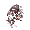
- PDB-2r5z: Structure of Scr/Exd complex bound to a DNA sequence derived from... -
+ Open data

- Basic information
| Entry | Database: PDB / ID: 2r5z | |||||||||
|---|---|---|---|---|---|---|---|---|---|---|
| Title | Structure of Scr/Exd complex bound to a DNA sequence derived from the fkh gene | |||||||||
 Components Components |
| |||||||||
 Keywords Keywords | TRANSCRIPTION/DNA /  Homeodomain / homeotic proteins / specificity / Developmental protein / DNA-binding / Homeobox / Nucleus / Transcription / Transcription regulation / Transcription-DNA COMPLEX Homeodomain / homeotic proteins / specificity / Developmental protein / DNA-binding / Homeobox / Nucleus / Transcription / Transcription regulation / Transcription-DNA COMPLEX | |||||||||
| Function / homology |  Function and homology information Function and homology informationsex comb development / salivary gland boundary specification / oenocyte development / somatic muscle development / eye development / regulation of cell fate specification / peripheral nervous system development / anterior/posterior pattern specification / midgut development / transcription factor binding ...sex comb development / salivary gland boundary specification / oenocyte development / somatic muscle development / eye development / regulation of cell fate specification / peripheral nervous system development / anterior/posterior pattern specification / midgut development / transcription factor binding / neuron development / embryonic organ development / cis-regulatory region sequence-specific DNA binding / protein-DNA complex / transcription coregulator activity / RNA polymerase II transcription regulatory region sequence-specific DNA binding / animal organ morphogenesis / brain development / RNA polymerase II transcription regulator complex / DNA-binding transcription factor binding / transcription regulator complex / sequence-specific DNA binding / transcription coactivator activity / DNA-binding transcription factor activity, RNA polymerase II-specific / RNA polymerase II cis-regulatory region sequence-specific DNA binding / DNA-binding transcription factor activity / protein heterodimerization activity / regulation of transcription by RNA polymerase II / positive regulation of DNA-templated transcription / protein homodimerization activity / positive regulation of transcription by RNA polymerase II / DNA binding / nucleoplasm / nucleus / cytoplasmSimilarity search - Function | |||||||||
| Biological species |  Drosophila melanogaster (fruit fly) Drosophila melanogaster (fruit fly)synthetic construct (others) | |||||||||
| Method | X-RAY DIFFRACTION / SYNCHROTRON / MOLECULAR REPLACEMENT / Resolution: 2.6 Å | |||||||||
 Authors Authors | Aggarwal, A.K. / Passner, J.M. / Jain, R. | |||||||||
 Citation Citation | Journal: Cell(Cambridge,Mass.) / Year: 2007 Title: Functional specificity of a Hox protein mediated by the recognition of minor groove structure Authors: Joshi, R. / Passner, J.M. / Rohs, R. / Jain, R. / Sosinsky, A. / Crickmore, M.A. / Jacob, V. / Aggarwal, A.K. / Honig, B. / Mann, R.S. | |||||||||
| History |
|
- Structure visualization
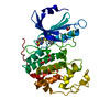
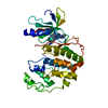
| Structure viewer | Molecule: MolmilJmol/JSmol |
|---|
- Downloads & links
-Download
| PDBx/mmCIF format | 2r5z.cif.gz | 68.3 KB | Display | PDBx/mmCIF format |
|---|---|---|---|---|
| PDB format | pdb2r5z.ent.gz | 46.9 KB | Display | PDB format |
| PDBx/mmJSON format | 2r5z.json.gz | Tree view | PDBx/mmJSON format | |
| Others |  Other downloads Other downloads |
-Validation report
| Arichive directory | https://data.pdbj.org/pub/pdb/validation_reports/r5/2r5zftp://data.pdbj.org/pub/pdb/validation_reports/r5/2r5z | HTTPS FTP |
|---|
-Related structure data
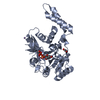
| Related structure data |  2r5yC  1b8iS S: Starting model for refinement C: citing same article ( |
|---|---|
| Similar structure data |


-Links
 PDBj
PDBj


- Assembly
| Deposited unit | 
| ||||||||
|---|---|---|---|---|---|---|---|---|---|
| 1 |
| ||||||||
| Unit cell |
|
-Components
| #1: DNA chain | Mass: 6132.991 Da / Num. of mol.: 1 / Source method: obtained synthetically / Source: (synth.) synthetic construct (others) / References: PDB-2R5Y |
|---|---|
| #2: DNA chain | Mass: 6132.991 Da / Num. of mol.: 1 / Source method: obtained synthetically / Source: (synth.) synthetic construct (others) / References: PDB-2R5Y |
| #3: Protein | Homeosis Mass: 10967.711 Da / Num. of mol.: 1 / Fragment: Homeobox DNA-binding domain Source method: isolated from a genetically manipulated source Source: (gene. exp.) Drosophila melanogaster (fruit fly) / Gene: Scr / Plasmid: pET-3A / Production host:  Escherichia coli (E. coli) / Strain (production host): BL21(DE3)pLysS / References: UniProt: P09077 Escherichia coli (E. coli) / Strain (production host): BL21(DE3)pLysS / References: UniProt: P09077 |
| #4: Protein | / Dpbx Mass: 7553.561 Da / Num. of mol.: 1 / Fragment: Homeobox TALE-type DNA-binding domain Source method: isolated from a genetically manipulated source Source: (gene. exp.) Drosophila melanogaster (fruit fly) / Gene: exd / Plasmid: pET-3A / Production host: Escherichia coli (E. coli) / Strain (production host): BL21(DE3)pLysS / References: UniProt: P40427 |
| #5: Water | ChemComp-HOH / Water Mass: 18.015 Da / Num. of mol.: 112 / Source method: isolated from a natural source / Formula: H2O Mass: 18.015 Da / Num. of mol.: 112 / Source method: isolated from a natural source / Formula: H2O |
-Experimental details
-Experiment
| Experiment | Method: X-RAY DIFFRACTION / Number of used crystals: 1 |
|---|
- Sample preparation
| Crystal | Density Matthews: 2.63 Å3/Da / Density % sol: 53.29 % |
|---|---|
| Crystal grow | pH: 8.7 Details: 10-14 % PEG 4K, 20 % MPD, 0.1 M Tris, pH 8.7-8.9, 0.2 M Sodium Acetate, 0.2 M Potassium Chloride |
-Data collection
| Diffraction | Mean temperature: 110 K |
|---|---|
| Diffraction source | Source: SYNCHROTRON / Site: APS  / Beamline: 19-ID / Wavelength: 1.1 Å / Beamline: 19-ID / Wavelength: 1.1 Å |
| Detector | Type: ADSC QUANTUM 315 / Detector: CCD / Date: Sep 10, 2004 Details: Rosenbaum-Rock high-resolution double-crystal monochromator. |
| Radiation | Monochromator: Rosenbaum-Rock high-resolution double-crystal monochromator. Protocol: SINGLE WAVELENGTH / Monochromatic (M) / Laue (L): M / Scattering type: x-ray |
| Radiation wavelength | Wavelength: 1.1 Å / Relative weight: 1 |
| Reflection | Resolution: 2.6→50 Å / Num. all: 10408 / Num. obs: 10189 / % possible obs: 97.9 % / Observed criterion σ(F): -3 / Redundancy: 33.6 % / Biso Wilson estimate: 41.7 Å2 / Rmerge(I) obs: 0.063 / Rsym value: 0.063 / Net I/σ(I): 36 |
| Reflection shell | Resolution: 2.6→2.69 Å / Redundancy: 1.86 % / Rmerge(I) obs: 0.32 / Mean I/σ(I) obs: 9.7 / Num. unique all: 1008 / Rsym value: 0.32 / % possible all: 100 |
- Processing
| Software |
| ||||||||||||||||||||||||||||||||||||||||||||||||||||||||||||||||||||||||||||||||
|---|---|---|---|---|---|---|---|---|---|---|---|---|---|---|---|---|---|---|---|---|---|---|---|---|---|---|---|---|---|---|---|---|---|---|---|---|---|---|---|---|---|---|---|---|---|---|---|---|---|---|---|---|---|---|---|---|---|---|---|---|---|---|---|---|---|---|---|---|---|---|---|---|---|---|---|---|---|---|---|---|---|
| Refinement | Method to determine structure: MOLECULAR REPLACEMENT Starting model: PDB entry 1B8I Resolution: 2.6→11.98 Å / Rfactor Rfree error: 0.013 / Data cutoff high absF: 1949550.74 / Data cutoff low absF: 0 / Isotropic thermal model: RESTRAINED / Cross valid method: THROUGHOUT / σ(F): 0 / Stereochemistry target values: Engh & Huber
| ||||||||||||||||||||||||||||||||||||||||||||||||||||||||||||||||||||||||||||||||
| Solvent computation | Solvent model: FLAT MODEL / Bsol: 45.4238 Å2 / ksol: 0.325294 e/Å3 | ||||||||||||||||||||||||||||||||||||||||||||||||||||||||||||||||||||||||||||||||
| Displacement parameters | Biso mean: 57.3 Å2
| ||||||||||||||||||||||||||||||||||||||||||||||||||||||||||||||||||||||||||||||||
| Refine analyze |
| ||||||||||||||||||||||||||||||||||||||||||||||||||||||||||||||||||||||||||||||||
| Refinement step | Cycle: LAST / Resolution: 2.6→11.98 Å
| ||||||||||||||||||||||||||||||||||||||||||||||||||||||||||||||||||||||||||||||||
| Refine LS restraints |
| ||||||||||||||||||||||||||||||||||||||||||||||||||||||||||||||||||||||||||||||||
| LS refinement shell | Resolution: 2.6→2.76 Å / Rfactor Rfree error: 0.042 / Total num. of bins used: 6
| ||||||||||||||||||||||||||||||||||||||||||||||||||||||||||||||||||||||||||||||||
| Xplor file |
|
