 Movie
Movie Controller

[English] 日本語
 Yorodumi
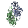
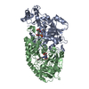
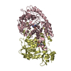
- PDB-2r0t: Crystal structure of GDP-4-keto-6-deoxymannose-3-dehydratase with... -
+ Open data

- Basic information
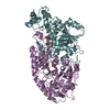
| Entry | Database: PDB / ID: 2r0t | ||||||
|---|---|---|---|---|---|---|---|
| Title | Crystal structure of GDP-4-keto-6-deoxymannose-3-dehydratase with a trapped PLP-glutamate geminal diamine | ||||||
 Components Components | Pyridoxamine 5-phosphate-dependent dehydrase | ||||||
 Keywords Keywords |  TRANSFERASE / colitose / aspartate aminotransferase / geminal diamine / pyridoxal phosphate TRANSFERASE / colitose / aspartate aminotransferase / geminal diamine / pyridoxal phosphate | ||||||
| Function / homology |  Function and homology informationTransferases; Transferring nitrogenous groups; Transaminases / transaminase activity Function and homology informationTransferases; Transferring nitrogenous groups; Transaminases / transaminase activitySimilarity search - Function | ||||||
| Biological species |  Escherichia coli (E. coli) Escherichia coli (E. coli) | ||||||
| Method | X-RAY DIFFRACTION / MOLECULAR REPLACEMENT / molecular replacement / Resolution: 1.9 Å | ||||||
 Authors Authors | Cook, P.D. / Holden, H.M. | ||||||
 Citation Citation | Journal: Biochemistry / Year: 2007 Title: A Structural Study of GDP-4-Keto-6-Deoxy-d-Mannose-3-Dehydratase: Caught in the Act of Geminal Diamine Formation Authors: Cook, P.D. / Holden, H.M. | ||||||
| History |
|
- Structure visualization
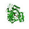
| Structure viewer | Molecule: MolmilJmol/JSmol |
|---|
- Downloads & links
-Download
| PDBx/mmCIF format | 2r0t.cif.gz | 169 KB | Display | PDBx/mmCIF format |
|---|---|---|---|---|
| PDB format | pdb2r0t.ent.gz | 132.8 KB | Display | PDB format |
| PDBx/mmJSON format | 2r0t.json.gz | Tree view | PDBx/mmJSON format | |
| Others |  Other downloads Other downloads |
-Validation report
| Arichive directory | https://data.pdbj.org/pub/pdb/validation_reports/r0/2r0tftp://data.pdbj.org/pub/pdb/validation_reports/r0/2r0t | HTTPS FTP |
|---|
-Related structure data
| Related structure data | 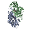 2gmuS S: Starting model for refinement |
|---|---|
| Similar structure data |


-Links
 PDBj
PDBj- Assembly
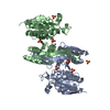
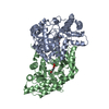
| Deposited unit | 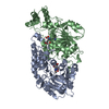
| ||||||||
|---|---|---|---|---|---|---|---|---|---|
| 1 |
| ||||||||
| Unit cell |
|
-Components
| #1: Protein | Mass: 44343.453 Da / Num. of mol.: 2 / Mutation: H188K Source method: isolated from a genetically manipulated source Source: (gene. exp.) Escherichia coli (E. coli) / Strain: O55:H7 Strain 5a / Gene: wbdK / Plasmid: pET28 / Production host: Escherichia coli (E. coli) / Strain (production host): Rosetta (DE3) / References: UniProt: Q9F118#2: Chemical | 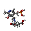  Mass: 378.272 Da / Num. of mol.: 2 / Source method: obtained synthetically / Formula: C13H19N2O9P Mass: 378.272 Da / Num. of mol.: 2 / Source method: obtained synthetically / Formula: C13H19N2O9P#3: Water | ChemComp-HOH / | Water Mass: 18.015 Da / Num. of mol.: 258 / Source method: isolated from a natural source / Formula: H2O Mass: 18.015 Da / Num. of mol.: 258 / Source method: isolated from a natural source / Formula: H2ONonpolymer details | LYSINE 188 IS COVALENTLY | |
|---|
-Experimental details
-Experiment
| Experiment | Method: X-RAY DIFFRACTION / Number of used crystals: 1 |
|---|
- Sample preparation
| Crystal | Density Matthews: 2.38 Å3/Da / Density % sol: 48.26 % |
|---|---|
| Crystal grow | Temperature: 298 K / pH: 6 Details: 24% PEG 3400, 100mM MES, 600mM KCl, 2mM PLP, 2mM 2-oxoglutarate, pH 6.0, batch, temperature 298K |
-Data collection
| Diffraction | Mean temperature: 298 K |
|---|---|
| Diffraction source | Source: ROTATING ANODE / Type: RIGAKU RU200 / Wavelength: 1.5418 |
| Detector | Type: BRUKER PROTEUM / Detector: CCD / Date: Jun 20, 2007 / Details: montel opics |
| Radiation | Monochromator: Ni filter / Protocol: SINGLE WAVELENGTH / Scattering type: x-ray |
| Radiation wavelength | Wavelength: 1.5418 Å / Relative weight: 1 |
| Reflection | Resolution: 1.9→30 Å / Num. all: 177105 / Num. obs: 60929 / % possible obs: 92.8 % / Observed criterion σ(F): 0 / Observed criterion σ(I): 0 / Redundancy: 2.9 % / Rsym value: 0.0626 / Net I/σ(I): 9.87 |
| Reflection shell | Resolution: 1.9→2 Å / Redundancy: 1.42 % / Mean I/σ(I) obs: 1.55 / Num. unique all: 7757 / Rsym value: 0.4 / % possible all: 83.3 |
-Phasing
| Phasing | Method: molecular replacement |
|---|
- Processing
| Software |
| ||||||||||||||||||||||||||||
|---|---|---|---|---|---|---|---|---|---|---|---|---|---|---|---|---|---|---|---|---|---|---|---|---|---|---|---|---|---|
| Refinement | Method to determine structure: MOLECULAR REPLACEMENT Starting model: PDB entry 2GMU Resolution: 1.9→30 Å / Isotropic thermal model: overall / Cross valid method: THROUGHOUT / σ(F): 0 / σ(I): 0 / Stereochemistry target values: Engh & Huber
| ||||||||||||||||||||||||||||
| Displacement parameters | Biso mean: 40.852 Å2 | ||||||||||||||||||||||||||||
| Refinement step | Cycle: LAST / Resolution: 1.9→30 Å
|
