 Movie
Movie Controller

[English] 日本語
 Yorodumi
- PDB-2qx3: Structure of pectate lyase II from Xanthomonas campestris pv. cam... -
+ Open data

- Basic information
| Entry | Database: PDB / ID: 2qx3 | ||||||
|---|---|---|---|---|---|---|---|
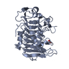
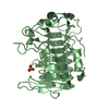
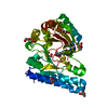
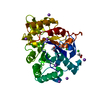
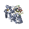
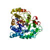
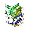
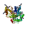
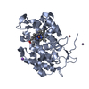
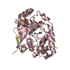
| Title | Structure of pectate lyase II from Xanthomonas campestris pv. campestris str. ATCC 33913 | ||||||
 Components Components | Pectate lyase II | ||||||
 Keywords Keywords | LYASE / parallel beta helix | ||||||
| Function / homology |  Function and homology informationpectate lyase activity / polysaccharide catabolic process / extracellular region Function and homology informationpectate lyase activity / polysaccharide catabolic process / extracellular regionSimilarity search - Function | ||||||
| Biological species |  Xanthomonas campestris pv. campestris (bacteria) Xanthomonas campestris pv. campestris (bacteria) | ||||||
| Method | X-RAY DIFFRACTION / MOLECULAR REPLACEMENT / Resolution: 2 Å | ||||||
 Authors Authors | Garron, M.L. / Shaya, D. | ||||||
 Citation Citation | Journal: To be Published Title: A seductive method to improve the thermostability and activity of an enzyme Authors: Lau, P.C. / Xiao, Z.Z. / Sulea, T. / Bergeron, H. / Grosse, S. / Beauchemin, M. | ||||||
| History |
|
- Structure visualization
| Structure viewer | Molecule: MolmilJmol/JSmol |
|---|
- Downloads & links
-Download
| PDBx/mmCIF format | 2qx3.cif.gz | 152.2 KB | Display | PDBx/mmCIF format |
|---|---|---|---|---|
| PDB format | pdb2qx3.ent.gz | 117.8 KB | Display | PDB format |
| PDBx/mmJSON format | 2qx3.json.gz | Tree view | PDBx/mmJSON format | |
| Others |  Other downloads Other downloads |
-Validation report
| Arichive directory | https://data.pdbj.org/pub/pdb/validation_reports/qx/2qx3ftp://data.pdbj.org/pub/pdb/validation_reports/qx/2qx3 | HTTPS FTP |
|---|
-Related structure data
| Related structure data |  1airS S: Starting model for refinement |
|---|---|
| Similar structure data |

-Links
 PDBj
PDBj


- Assembly
| Deposited unit | 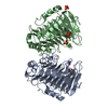
| ||||||||
|---|---|---|---|---|---|---|---|---|---|
| 1 | 
| ||||||||
| 2 | 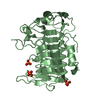
| ||||||||
| Unit cell |
|
-Components
| #1: Protein | Mass: 35275.082 Da / Num. of mol.: 2 Source method: isolated from a genetically manipulated source Source: (gene. exp.) Xanthomonas campestris pv. campestris (bacteria)Species: Xanthomonas campestris / Strain: ATCC 33913 / Gene: pelB / Production host: Escherichia coli (E. coli) / References: UniProt: Q8P6Z9, pectate lyase#2: Chemical | ChemComp-PO4 / Phosphate  Mass: 94.971 Da / Num. of mol.: 5 / Source method: obtained synthetically / Formula: PO4 Mass: 94.971 Da / Num. of mol.: 5 / Source method: obtained synthetically / Formula: PO4#3: Water | ChemComp-HOH / | Water Mass: 18.015 Da / Num. of mol.: 598 / Source method: isolated from a natural source / Formula: H2O Mass: 18.015 Da / Num. of mol.: 598 / Source method: isolated from a natural source / Formula: H2O |
|---|
-Experimental details
-Experiment
| Experiment | Method: X-RAY DIFFRACTION / Number of used crystals: 1 |
|---|
- Sample preparation
| Crystal | Density Matthews: 2.3 Å3/Da / Density % sol: 46.48 % |
|---|---|
| Crystal grow | Temperature: 294 K / Method: vapor diffusion, hanging drop / pH: 7 Details: 0.1M Hepes, 25% polyethylene glycol 3350, pH 7, VAPOR DIFFUSION, HANGING DROP, temperature 294K |
-Data collection
| Diffraction | Mean temperature: 100 K | |||||||||||||||||||||||||||||||||||||||||||||||||||||||||||||||||||||||||||||
|---|---|---|---|---|---|---|---|---|---|---|---|---|---|---|---|---|---|---|---|---|---|---|---|---|---|---|---|---|---|---|---|---|---|---|---|---|---|---|---|---|---|---|---|---|---|---|---|---|---|---|---|---|---|---|---|---|---|---|---|---|---|---|---|---|---|---|---|---|---|---|---|---|---|---|---|---|---|---|
| Diffraction source | Source: ROTATING ANODE / Type: RIGAKU MICROMAX-007 / Wavelength: 1.5418 | |||||||||||||||||||||||||||||||||||||||||||||||||||||||||||||||||||||||||||||
| Detector | Type: RIGAKU RAXIS HTC / Detector: IMAGE PLATE / Date: May 1, 2007 | |||||||||||||||||||||||||||||||||||||||||||||||||||||||||||||||||||||||||||||
| Radiation | Protocol: SINGLE WAVELENGTH / Monochromatic (M) / Laue (L): M / Scattering type: x-ray | |||||||||||||||||||||||||||||||||||||||||||||||||||||||||||||||||||||||||||||
| Radiation wavelength | Wavelength: 1.5418 Å / Relative weight: 1 | |||||||||||||||||||||||||||||||||||||||||||||||||||||||||||||||||||||||||||||
| Reflection | Redundancy: 1.9 % / Av σ(I) over netI: 20.1 / Number: 66970 / Rmerge(I) obs: 0.058 / Χ2: 3.02 / D res high: 2 Å / D res low: 25 Å / Num. obs: 35612 / % possible obs: 83.6 | |||||||||||||||||||||||||||||||||||||||||||||||||||||||||||||||||||||||||||||
| Diffraction reflection shell |
| |||||||||||||||||||||||||||||||||||||||||||||||||||||||||||||||||||||||||||||
| Reflection | Resolution: 2→25 Å / Num. all: 35612 / Num. obs: 35612 / % possible obs: 83.6 % / Observed criterion σ(F): 0 / Observed criterion σ(I): 0 / Redundancy: 1.9 % / Rmerge(I) obs: 0.058 / Χ2: 3.024 / Net I/σ(I): 20.1 | |||||||||||||||||||||||||||||||||||||||||||||||||||||||||||||||||||||||||||||
| Reflection shell |
|
-Phasing
| Phasing MR |
|
|---|
- Processing
| Software |
| ||||||||||||||||||||||||||||||||||||||||||||||||||||||||||||||||||||||||||||||||||||||||||
|---|---|---|---|---|---|---|---|---|---|---|---|---|---|---|---|---|---|---|---|---|---|---|---|---|---|---|---|---|---|---|---|---|---|---|---|---|---|---|---|---|---|---|---|---|---|---|---|---|---|---|---|---|---|---|---|---|---|---|---|---|---|---|---|---|---|---|---|---|---|---|---|---|---|---|---|---|---|---|---|---|---|---|---|---|---|---|---|---|---|---|---|
| Refinement | Method to determine structure: MOLECULAR REPLACEMENT Starting model: PDB ENTRY 1AIR Resolution: 2→25 Å / Cor.coef. Fo:Fc: 0.964 / Cor.coef. Fo:Fc free: 0.937 / SU B: 4.088 / SU ML: 0.116 / Cross valid method: THROUGHOUT / σ(F): 0 / ESU R: 0.252 / ESU R Free: 0.189 / Stereochemistry target values: MAXIMUM LIKELIHOOD / Details: HYDROGENS HAVE BEEN ADDED IN THE RIDING POSITIONS
| ||||||||||||||||||||||||||||||||||||||||||||||||||||||||||||||||||||||||||||||||||||||||||
| Solvent computation | Ion probe radii: 0.8 Å / Shrinkage radii: 0.8 Å / VDW probe radii: 1.4 Å / Solvent model: BABINET MODEL WITH MASK | ||||||||||||||||||||||||||||||||||||||||||||||||||||||||||||||||||||||||||||||||||||||||||
| Displacement parameters | Biso mean: 23.302 Å2
| ||||||||||||||||||||||||||||||||||||||||||||||||||||||||||||||||||||||||||||||||||||||||||
| Refinement step | Cycle: LAST / Resolution: 2→25 Å
| ||||||||||||||||||||||||||||||||||||||||||||||||||||||||||||||||||||||||||||||||||||||||||
| Refine LS restraints |
| ||||||||||||||||||||||||||||||||||||||||||||||||||||||||||||||||||||||||||||||||||||||||||
| LS refinement shell | Resolution: 2→2.047 Å / Total num. of bins used: 20
|
