- PDB-2qrv: Structure of Dnmt3a-Dnmt3L C-terminal domain complex -
+
Open data
ID or keywords:
Loading...
-
Basic information
Entry
Database: PDB / ID: 2qrv
Title
Structure of Dnmt3a-Dnmt3L C-terminal domain complex
Components
DNA (cytosine-5)-methyltransferase 3-like
DNA (cytosine-5)-methyltransferase 3ADNA (cytosine-5)-methyltransferase 3A
Keywords
transferase/transferase regulator / DNA methyltransferase 3a (Dnmt3a) and its regulatory factor / DNA methyltransferase 3-like protein (Dnmt3L) / Nucleus / S-adenosyl-L-methionine / transferase-transferase regulator COMPLEX
Function / homology
Function and homology information
retrotransposon silencing by heterochromatin formation / epigenetic programing of female pronucleus / chorionic trophoblast cell differentiation / positive regulation of cellular response to hypoxia / DNA (cytosine-5-)-methyltransferase activity, acting on CpG substrates / epigenetic programming of gene expression / cellular response to bisphenol A / protein-cysteine methyltransferase activity / genomic imprinting / DNA (cytosine-5-)-methyltransferase ...retrotransposon silencing by heterochromatin formation / epigenetic programing of female pronucleus / chorionic trophoblast cell differentiation / positive regulation of cellular response to hypoxia / DNA (cytosine-5-)-methyltransferase activity, acting on CpG substrates / epigenetic programming of gene expression / cellular response to bisphenol A / protein-cysteine methyltransferase activity / genomic imprinting / DNA (cytosine-5-)-methyltransferase / unmethylated CpG binding / DNA (cytosine-5-)-methyltransferase activity / autosome genomic imprinting / S-adenosylmethionine metabolic process / negative regulation of DNA methylation-dependent heterochromatin formation / SUMOylation of DNA methylation proteins / DNA methylation-dependent heterochromatin formation / ESC/E(Z) complex / XY body / cellular response to ethanol / response to vitamin A / negative regulation of gene expression via chromosomal CpG island methylation / DNA metabolic process / negative regulation of gene expression, epigenetic / response to ionizing radiation / hepatocyte apoptotic process / male meiosis I / chromosome, centromeric region / catalytic complex / heterochromatin / enzyme activator activity / DNA methylation / Transferases; Transferring one-carbon groups; Methyltransferases / PRC2 methylates histones and DNA / response to cocaine / condensed nuclear chromosome / Defective pyroptosis / stem cell differentiation / placenta development / cellular response to amino acid stimulus / response to lead ion / euchromatin / neuron differentiation / response to toxic substance / RMTs methylate histone arginines / nuclear matrix / transcription corepressor activity / response to estradiol / cellular response to hypoxia / methylation / spermatogenesis / RNA polymerase II-specific DNA-binding transcription factor binding / response to xenobiotic stimulus / RNA polymerase II cis-regulatory region sequence-specific DNA binding / negative regulation of DNA-templated transcription / chromatin binding / enzyme binding / negative regulation of transcription by RNA polymerase II / DNA binding / nucleoplasm / identical protein binding / metal ion binding / nucleus / cytosol / cytoplasm Similarity search - Function
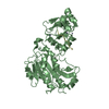
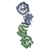
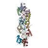
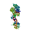
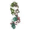
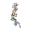
A: DNA (cytosine-5)-methyltransferase 3A B: DNA (cytosine-5)-methyltransferase 3-like C: DNA (cytosine-5)-methyltransferase 3-like D: DNA (cytosine-5)-methyltransferase 3A E: DNA (cytosine-5)-methyltransferase 3A F: DNA (cytosine-5)-methyltransferase 3-like G: DNA (cytosine-5)-methyltransferase 3-like H: DNA (cytosine-5)-methyltransferase 3A hetero molecules
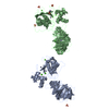
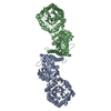
A: DNA (cytosine-5)-methyltransferase 3A B: DNA (cytosine-5)-methyltransferase 3-like C: DNA (cytosine-5)-methyltransferase 3-like D: DNA (cytosine-5)-methyltransferase 3A hetero molecules
E: DNA (cytosine-5)-methyltransferase 3A F: DNA (cytosine-5)-methyltransferase 3-like G: DNA (cytosine-5)-methyltransferase 3-like H: DNA (cytosine-5)-methyltransferase 3A hetero molecules
There were two tetramer complexes per crystallographic asymmetric unit. Each tetramer contains two pairs of Dnmt3a-3L hetero-dimers (3L-3a-3a-3L) with two 3L-3a interfaces and one 3a-3a interface.
-
Components
#1: Protein
DNA (cytosine-5)-methyltransferase3A / DNA (cytosine-5)-methyltransferase 3A / Dnmt3a / DNA methyltransferase HsaIIIA / DNA MTase HsaIIIA / M.HsaIIIA
Mass: 33961.148 Da / Num. of mol.: 4 / Fragment: residues 623 to 908 Source method: isolated from a genetically manipulated source Source: (gene. exp.) Homo sapiens (human) / Gene: DNMT3A / Plasmid: pXC528 / Species (production host): Escherichia coli / Production host: Escherichia coli BL21(DE3) (bacteria) / Strain (production host): BL21 (DE3) References: UniProt: Q9Y6K1, DNA (cytosine-5-)-methyltransferase
#2: Protein
DNA (cytosine-5)-methyltransferase3-like
Mass: 26692.486 Da / Num. of mol.: 4 / Fragment: residues 160 to 386 Source method: isolated from a genetically manipulated source Source: (gene. exp.) Homo sapiens (human) / Gene: DNMT3L / Plasmid: pXC510 / Species (production host): Escherichia coli / Production host: Escherichia coli BL21(DE3) (bacteria) / Strain (production host): BL21 (DE3) / References: UniProt: Q9UJW3
Type: L-peptide linking / Mass: 384.411 Da / Num. of mol.: 4 / Source method: obtained synthetically / Formula: C14H20N6O5S
-
Experimental details
-
Experiment
Experiment
Method: X-RAY DIFFRACTION / Number of used crystals: 1
-
Sample preparation
Crystal
Density Matthews: 3.18 Å3/Da / Density % sol: 61.29 % Description: THERE ARE TWO TETRAMER COMPLEXES PER ASYMMETRIC UNIT. TETRAMER FORMED BY CHAINS A,B,C,D IS MORE STABLE THAN THE TETRAMER FORMED BY CHAINS E,F,G,H. WITHIN EACH TETRAMER, THE DNMT3L ...Description: THERE ARE TWO TETRAMER COMPLEXES PER ASYMMETRIC UNIT. TETRAMER FORMED BY CHAINS A,B,C,D IS MORE STABLE THAN THE TETRAMER FORMED BY CHAINS E,F,G,H. WITHIN EACH TETRAMER, THE DNMT3L MOLECULES HAVE HIGHER B-FACTOR THAN THAT OF DNMT3A, INDICATING DNMT3L MOLECULES - LOCATED IN THE OUTER SURFACES OF TETRAMER - ARE MORE MOBILE.
Crystal grow
Temperature: 300 K / Method: vapor diffusion, hanging drop / pH: 8 Details: The final protein solution CONTAINED ~100 TO 133 micro M COMPLEX IN 20 milli M TRIS/HCL, PH 8.0, 100 milli M NACL, 5 % GLYCEROL, AND 0.1 % MERCAPTOETHANOL. Crystals were obtained with the ...Details: The final protein solution CONTAINED ~100 TO 133 micro M COMPLEX IN 20 milli M TRIS/HCL, PH 8.0, 100 milli M NACL, 5 % GLYCEROL, AND 0.1 % MERCAPTOETHANOL. Crystals were obtained with the mother liquor containing 2~5 % PEG 3000, 100 mM Tris/HCl, pH 8.0, 5 % glycerol at 16C, VAPOR DIFFUSION, HANGING DROP
Resolution: 2.89→38.9 Å / Rfactor Rfree error: 0.005 / Data cutoff high absF: 332407.34 / Data cutoff high rms absF: 332407.34 / Data cutoff low absF: 0 / Isotropic thermal model: RESTRAINED / Cross valid method: THROUGHOUT / σ(F): 2 / Stereochemistry target values: ENGH & HUBER Details: The non-crystallographic symmetry was used as restrains in the CNS refinement, and was released for the side chains at the later cycles to account for different interaction environments of ...Details: The non-crystallographic symmetry was used as restrains in the CNS refinement, and was released for the side chains at the later cycles to account for different interaction environments of crystal packing with each molecule. The group B-factor for each residue was refined at the earlier stage of refinement and the individual B-factor for each atom was refined at the later cycles.
In the structure databanks used in Yorodumi, some data are registered as the other names, "COVID-19 virus" and "2019-nCoV". Here are the details of the virus and the list of structure data.
Jan 31, 2019. EMDB accession codes are about to change! (news from PDBe EMDB page)
EMDB accession codes are about to change! (news from PDBe EMDB page)
The allocation of 4 digits for EMDB accession codes will soon come to an end. Whilst these codes will remain in use, new EMDB accession codes will include an additional digit and will expand incrementally as the available range of codes is exhausted. The current 4-digit format prefixed with “EMD-” (i.e. EMD-XXXX) will advance to a 5-digit format (i.e. EMD-XXXXX), and so on. It is currently estimated that the 4-digit codes will be depleted around Spring 2019, at which point the 5-digit format will come into force.
The EM Navigator/Yorodumi systems omit the EMD- prefix.
Related info.:Q: What is EMD? / ID/Accession-code notation in Yorodumi/EM Navigator
Yorodumi is a browser for structure data from EMDB, PDB, SASBDB, etc.
This page is also the successor to EM Navigator detail page, and also detail information page/front-end page for Omokage search.
The word "yorodu" (or yorozu) is an old Japanese word meaning "ten thousand". "mi" (miru) is to see.
Related info.:EMDB / PDB / SASBDB / Comparison of 3 databanks / Yorodumi Search / Aug 31, 2016. New EM Navigator & Yorodumi / Yorodumi Papers / Jmol/JSmol / Function and homology information / Changes in new EM Navigator and Yorodumi
 Movie
Movie Controller


 Open data
 Basic information
 Components
 Keywords
 Nucleus /
  Function and homology information
 Authors
 Citation
 Structure visualization
 Downloads & links
 Other downloads
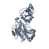


 PDBj
PDBj

 Assembly
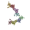


 Type: L-peptide linking / Mass: 384.411 Da / Num. of mol.: 4 / Source method: obtained synthetically / Formula: C14H20N6O5S
 Sample preparation
 / Beamline: 22-ID / Wavelength: 1.0, 0.9792, 0.9785,0.9719
 Processing