 Movie
Movie Controller


+ Open data

- Basic information
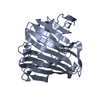
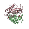
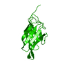
| Entry | Database: PDB / ID: 2qdn | ||||||
|---|---|---|---|---|---|---|---|
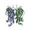
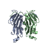
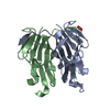
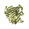
| Title | Crystal Structure of mouse GITRL | ||||||
 Components Components | GITR ligand | ||||||
 Keywords Keywords |  IMMUNE SYSTEM / GITRL / Glucocorticoid-Induced TNF Receptor Ligand IMMUNE SYSTEM / GITRL / Glucocorticoid-Induced TNF Receptor Ligand | ||||||
| Function / homology |  Function and homology information Function and homology informationregulation of dendritic cell chemotaxis / negative regulation of T-helper 17 cell lineage commitment / TNFs bind their physiological receptors / tumor necrosis factor receptor superfamily binding / tumor necrosis factor receptor binding / positive regulation of monocyte chemotaxis / positive regulation of leukocyte migration / positive regulation of macrophage chemotaxis / regulation of T cell proliferation / T cell proliferation involved in immune response ...regulation of dendritic cell chemotaxis / negative regulation of T-helper 17 cell lineage commitment / TNFs bind their physiological receptors / tumor necrosis factor receptor superfamily binding / tumor necrosis factor receptor binding / positive regulation of monocyte chemotaxis / positive regulation of leukocyte migration / positive regulation of macrophage chemotaxis / regulation of T cell proliferation / T cell proliferation involved in immune response / positive regulation of cell adhesion / regulation of protein-containing complex assembly / positive regulation of tyrosine phosphorylation of STAT protein / tumor necrosis factor-mediated signaling pathway / cytokine activity / positive regulation of inflammatory response / positive regulation of NF-kappaB transcription factor activity / adaptive immune response / cell surface / extracellular space / identical protein binding / plasma membraneSimilarity search - Function | ||||||
| Biological species |  Mus musculus (house mouse) Mus musculus (house mouse) | ||||||
| Method | X-RAY DIFFRACTION / SYNCHROTRON / SAD / Resolution: 2.09 Å | ||||||
 Authors Authors | Chattopadhyay, K. / Ramagopal, U.A. / Nathenson, S.G. / Almo, S.C. | ||||||
 Citation Citation | Journal: Proc.Natl.Acad.Sci.Usa / Year: 2008 Title: Evolution of GITRL immune function: murine GITRL exhibits unique structural and biochemical properties within the TNF superfamily. Authors: Chattopadhyay, K. / Ramagopal, U.A. / Brenowitz, M. / Nathenson, S.G. / Almo, S.C. | ||||||
| History |
|
- Structure visualization
| Structure viewer | Molecule: MolmilJmol/JSmol |
|---|
- Downloads & links
-Download
| PDBx/mmCIF format | 2qdn.cif.gz | 62.8 KB | Display | PDBx/mmCIF format |
|---|---|---|---|---|
| PDB format | pdb2qdn.ent.gz | 49.8 KB | Display | PDB format |
| PDBx/mmJSON format | 2qdn.json.gz | Tree view | PDBx/mmJSON format | |
| Others |  Other downloads Other downloads |
-Validation report
| Arichive directory | https://data.pdbj.org/pub/pdb/validation_reports/qd/2qdnftp://data.pdbj.org/pub/pdb/validation_reports/qd/2qdn | HTTPS FTP |
|---|
-Related structure data


-Links
 PDBj
PDBj- Assembly
| Deposited unit | 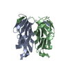
| ||||||||
|---|---|---|---|---|---|---|---|---|---|
| 1 |
| ||||||||
| Unit cell |
|
-Components
| #1: Protein | Mass: 15011.407 Da / Num. of mol.: 2 / Fragment: TNF homology domain (Residues 46-173) Source method: isolated from a genetically manipulated source Source: (gene. exp.) Mus musculus (house mouse) / Gene: Gitrl / Plasmid: pET-14b / Production host:  Escherichia coli (E. coli) / Strain (production host): Rosetta (DE3) pLysS / References: UniProt: Q7TS55 Escherichia coli (E. coli) / Strain (production host): Rosetta (DE3) pLysS / References: UniProt: Q7TS55#2: Water | ChemComp-HOH / | Water Mass: 18.015 Da / Num. of mol.: 223 / Source method: isolated from a natural source / Formula: H2O Mass: 18.015 Da / Num. of mol.: 223 / Source method: isolated from a natural source / Formula: H2O |
|---|
-Experimental details
-Experiment
| Experiment | Method: X-RAY DIFFRACTION / Number of used crystals: 1 |
|---|
- Sample preparation
| Crystal | Density Matthews: 2.21 Å3/Da / Density % sol: 44.36 % |
|---|---|
| Crystal grow | Temperature: 298 K / Method: vapor diffusion, sitting drop / pH: 7 Details: 15% PEG 3350, 0.1 M Succinic acid pH 7.0, Vapor diffusion, Sitting drop, temperature 298K |
-Data collection
| Diffraction | Mean temperature: 100 K | |||||||||||||||||||||||||||||||||||||||||||||||||||||||||||||||||||||||||||||
|---|---|---|---|---|---|---|---|---|---|---|---|---|---|---|---|---|---|---|---|---|---|---|---|---|---|---|---|---|---|---|---|---|---|---|---|---|---|---|---|---|---|---|---|---|---|---|---|---|---|---|---|---|---|---|---|---|---|---|---|---|---|---|---|---|---|---|---|---|---|---|---|---|---|---|---|---|---|---|
| Diffraction source | Source: SYNCHROTRON / Site: NSLS  / Beamline: X4A / Wavelength: 1.74332 Å / Beamline: X4A / Wavelength: 1.74332 Å | |||||||||||||||||||||||||||||||||||||||||||||||||||||||||||||||||||||||||||||
| Detector | Type: ADSC QUANTUM 4 / Detector: CCD / Date: May 1, 2006 | |||||||||||||||||||||||||||||||||||||||||||||||||||||||||||||||||||||||||||||
| Radiation | Protocol: SINGLE WAVELENGTH / Monochromatic (M) / Laue (L): M / Scattering type: x-ray | |||||||||||||||||||||||||||||||||||||||||||||||||||||||||||||||||||||||||||||
| Radiation wavelength | Wavelength: 1.74332 Å / Relative weight: 1 | |||||||||||||||||||||||||||||||||||||||||||||||||||||||||||||||||||||||||||||
| Reflection | Redundancy: 11.2 % / Av σ(I) over netI: 27.3 / Number: 309693 / Rmerge(I) obs: 0.043 / Χ2: 1.15 / D res high: 2.09 Å / D res low: 50 Å / Num. obs: 27694 / % possible obs: 90.3 | |||||||||||||||||||||||||||||||||||||||||||||||||||||||||||||||||||||||||||||
| Diffraction reflection shell |
| |||||||||||||||||||||||||||||||||||||||||||||||||||||||||||||||||||||||||||||
| Reflection | Resolution: 2.09→50.06 Å / Num. all: 14872 / Num. obs: 14872 / % possible obs: 90.3 % / Observed criterion σ(F): 0 / Observed criterion σ(I): 0 / Redundancy: 11.2 % / Rmerge(I) obs: 0.043 / Rsym value: 0.048 / Χ2: 1.154 / Net I/σ(I): 27.3 | |||||||||||||||||||||||||||||||||||||||||||||||||||||||||||||||||||||||||||||
| Reflection shell |
|
- Processing
| Software |
| |||||||||||||||||||||||||||||||||||||||||||||||||||||||||||||||||||||||||||||||||||||
|---|---|---|---|---|---|---|---|---|---|---|---|---|---|---|---|---|---|---|---|---|---|---|---|---|---|---|---|---|---|---|---|---|---|---|---|---|---|---|---|---|---|---|---|---|---|---|---|---|---|---|---|---|---|---|---|---|---|---|---|---|---|---|---|---|---|---|---|---|---|---|---|---|---|---|---|---|---|---|---|---|---|---|---|---|---|---|
| Refinement | Method to determine structure: SAD / Resolution: 2.09→50.06 Å / Cor.coef. Fo:Fc: 0.951 / Cor.coef. Fo:Fc free: 0.923 / SU B: 4.305 / SU ML: 0.119 / Cross valid method: THROUGHOUT / σ(F): 0 / σ(I): 0 / ESU R: 0.254 / ESU R Free: 0.201 / Stereochemistry target values: MAXIMUM LIKELIHOOD / Details: HYDROGENS HAVE BEEN ADDED IN THE RIDING POSITIONS
| |||||||||||||||||||||||||||||||||||||||||||||||||||||||||||||||||||||||||||||||||||||
| Solvent computation | Ion probe radii: 0.8 Å / Shrinkage radii: 0.8 Å / VDW probe radii: 1.2 Å / Solvent model: MASK | |||||||||||||||||||||||||||||||||||||||||||||||||||||||||||||||||||||||||||||||||||||
| Displacement parameters | Biso mean: 21.558 Å2
| |||||||||||||||||||||||||||||||||||||||||||||||||||||||||||||||||||||||||||||||||||||
| Refinement step | Cycle: LAST / Resolution: 2.09→50.06 Å
| |||||||||||||||||||||||||||||||||||||||||||||||||||||||||||||||||||||||||||||||||||||
| Refine LS restraints |
| |||||||||||||||||||||||||||||||||||||||||||||||||||||||||||||||||||||||||||||||||||||
| LS refinement shell | Resolution: 2.09→2.139 Å / Total num. of bins used: 20
|
