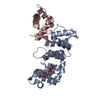
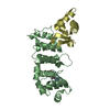
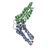
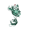
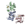
Entry Database : PDB / ID : 2qdjTitle Crystal structure of the Retinoblastoma protein N-domain provides insight into tumor suppression, ligand interaction and holoprotein architecture Retinoblastoma-associated protein Keywords / / Function / homology Function Domain/homology Component
/ / / / / / / / / / / / / / / / / / / / / / / / / / / / / / / / / / / / / / / / / / / / / / / / / / / / / / / / / / / / / / / / / / / / / / / / / / / / / / / / / / / / / / / / / / / / / / / / / / / / / / / / / / / / / / / / / / / / / / / / / / / / / / / / / / / / / / / Biological species Homo sapiens (human)Method / / / Resolution : 2 Å Authors Hassler, M. / Mittnacht, S. / Pearl, L.H. Journal : Mol.Cell / Year : 2007Title : Crystal structure of the retinoblastoma protein N domain provides insight into tumor suppression, ligand interaction, and holoprotein architecture.Authors : Hassler, M. / Singh, S. / Yue, W.W. / Luczynski, M. / Lakbir, R. / Sanchez-Sanchez, F. / Bader, T. / Pearl, L.H. / Mittnacht, S. History Deposition Jun 21, 2007 Deposition site / Processing site Revision 1.0 Jan 22, 2008 Provider / Type Revision 1.1 Jul 13, 2011 Group
Show all Show less
 Movie
Movie Controller

 Yorodumi
 Open data
 Basic information
 Components
 Keywords
 ANTITUMOR PROTEIN / cyclin fold / cyclin wedge
 Function and homology information
 Authors
 Citation
 Structure visualization
 Downloads & links
 Other downloads
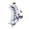


 PDBj
PDBj


 Assembly

 Mass: 18.015 Da / Num. of mol.: 234 / Source method: isolated from a natural source / Formula: H2O
 Sample preparation
 / Beamline: ID14-2 / Wavelength: 0.933 Å
 Processing