 Movie
Movie Controller

[English] 日本語
 Yorodumi
- PDB-2q9x: Crystal structure of highly stable mutant Q40P/S47I/H93G of human... -
+ Open data

- Basic information
| Entry | Database: PDB / ID: 2q9x | ||||||
|---|---|---|---|---|---|---|---|
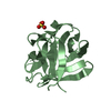
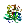
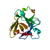
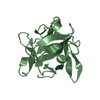
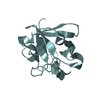
| Title | Crystal structure of highly stable mutant Q40P/S47I/H93G of human fibroblast growth factor-1 | ||||||
 Components Components | Heparin-binding growth factor 1 | ||||||
 Keywords Keywords |  HORMONE / FGF-1 / growth factor / beta trefoil / enhanced stability HORMONE / FGF-1 / growth factor / beta trefoil / enhanced stability | ||||||
| Function / homology |  Function and homology information Function and homology informationmesonephric epithelium development / branch elongation involved in ureteric bud branching / regulation of endothelial tube morphogenesis / FGFR3b ligand binding and activation / regulation of endothelial cell chemotaxis to fibroblast growth factor / Signaling by activated point mutants of FGFR3 / FGFR3c ligand binding and activation / Phospholipase C-mediated cascade; FGFR3 / FGFR2b ligand binding and activation / positive regulation of cholesterol biosynthetic process ...mesonephric epithelium development / branch elongation involved in ureteric bud branching / regulation of endothelial tube morphogenesis / FGFR3b ligand binding and activation / regulation of endothelial cell chemotaxis to fibroblast growth factor / Signaling by activated point mutants of FGFR3 / FGFR3c ligand binding and activation / Phospholipase C-mediated cascade; FGFR3 / FGFR2b ligand binding and activation / positive regulation of cholesterol biosynthetic process / fibroblast growth factor receptor binding / FGFR2c ligand binding and activation / Activated point mutants of FGFR2 / Phospholipase C-mediated cascade; FGFR2 / FGFR4 ligand binding and activation / FGFR1b ligand binding and activation / Phospholipase C-mediated cascade; FGFR4 / Signaling by activated point mutants of FGFR1 / FGFR1c ligand binding and activation / organ induction / Downstream signaling of activated FGFR1 / Phospholipase C-mediated cascade: FGFR1 / positive regulation of hepatocyte proliferation / S100 protein binding / positive regulation of intracellular signal transduction / Signaling by FGFR2 IIIa TM / PI-3K cascade:FGFR3 / positive regulation of sprouting angiogenesis / PI-3K cascade:FGFR2 / PI-3K cascade:FGFR4 / PI-3K cascade:FGFR1 / positive regulation of cell division / PI3K Cascade / anatomical structure morphogenesis / fibroblast growth factor receptor signaling pathway / SHC-mediated cascade:FGFR3 / SHC-mediated cascade:FGFR2 / SHC-mediated cascade:FGFR4 / SHC-mediated cascade:FGFR1 / FRS-mediated FGFR3 signaling / FRS-mediated FGFR2 signaling / FRS-mediated FGFR4 signaling / Signaling by FGFR3 in disease / FRS-mediated FGFR1 signaling / Hsp70 protein binding / Signaling by FGFR2 in disease / Signaling by FGFR1 in disease / activation of protein kinase B activity / extracellular matrix / positive regulation of endothelial cell migration / epithelial cell proliferation / positive regulation of epithelial cell proliferation / Negative regulation of FGFR3 signaling / Negative regulation of FGFR2 signaling / Negative regulation of FGFR4 signaling / Negative regulation of FGFR1 signaling / animal organ morphogenesis / lung development / growth factor activity / positive regulation of MAP kinase activity / wound healing / positive regulation of angiogenesis / Constitutive Signaling by Aberrant PI3K in Cancer / integrin binding / PIP3 activates AKT signaling / cellular response to heat / heparin binding / cell cortex / PI5P, PP2A and IER3 Regulate PI3K/AKT Signaling / RAF/MAP kinase cascade / angiogenesis / cell differentiation / positive regulation of ERK1 and ERK2 cascade / positive regulation of cell migration / positive regulation of cell population proliferation / positive regulation of gene expression / signal transduction / positive regulation of transcription by RNA polymerase II / extracellular space / extracellular region / nucleoplasm / nucleus / cytosol / cytoplasmSimilarity search - Function | ||||||
| Biological species |  Homo sapiens (human) Homo sapiens (human) | ||||||
| Method | X-RAY DIFFRACTION / SYNCHROTRON / MOLECULAR REPLACEMENT / Resolution: 1.7 Å | ||||||
 Authors Authors | Szlachcic, A. / Zakrzewska, M. / Krowarsch, D. / Os, V. / Helland, R. / Otlewski, J. | ||||||
 Citation Citation | Journal: To be Published Title: Crystal structure of highly stable mutant Q40P/S47I/H93G of human fibroblast growth factor-1 Authors: Szlachcic, A. / Zakrzewska, M. / Krowarsch, D. / Os, V. / Helland, R. / Otlewski, J. | ||||||
| History |
|
- Structure visualization
| Structure viewer | Molecule: MolmilJmol/JSmol |
|---|
- Downloads & links
-Download
| PDBx/mmCIF format | 2q9x.cif.gz | 41.7 KB | Display | PDBx/mmCIF format |
|---|---|---|---|---|
| PDB format | pdb2q9x.ent.gz | 28 KB | Display | PDB format |
| PDBx/mmJSON format | 2q9x.json.gz | Tree view | PDBx/mmJSON format | |
| Others |  Other downloads Other downloads |
-Validation report
| Arichive directory | https://data.pdbj.org/pub/pdb/validation_reports/q9/2q9xftp://data.pdbj.org/pub/pdb/validation_reports/q9/2q9x | HTTPS FTP |
|---|
-Related structure data
| Related structure data |  1rg8S S: Starting model for refinement |
|---|---|
| Similar structure data |

-Links
 PDBj
PDBj


- Assembly
| Deposited unit | 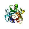
| ||||||||
|---|---|---|---|---|---|---|---|---|---|
| 1 |
| ||||||||
| Unit cell |
| ||||||||
| Details | BIOLOGICAL ASSEMBLY IS A MONOMER |
-Components
| #1: Protein | Mass: 15771.837 Da / Num. of mol.: 1 / Mutation: Q40P, S47I, H93G Source method: isolated from a genetically manipulated source Source: (gene. exp.) Homo sapiens (human) / Gene: FGF1, FGFA / Plasmid: pET3c / Production host:  Escherichia coli (E. coli) / Strain (production host): BL21(DE3)pLysS / References: UniProt: P05230 Escherichia coli (E. coli) / Strain (production host): BL21(DE3)pLysS / References: UniProt: P05230 |
|---|---|
| #2: Chemical | ChemComp-GOL / Glycerol  Mass: 92.094 Da / Num. of mol.: 1 / Source method: obtained synthetically / Formula: C3H8O3 Mass: 92.094 Da / Num. of mol.: 1 / Source method: obtained synthetically / Formula: C3H8O3 |
| #3: Water | ChemComp-HOH / Water Mass: 18.015 Da / Num. of mol.: 107 / Source method: isolated from a natural source / Formula: H2O Mass: 18.015 Da / Num. of mol.: 107 / Source method: isolated from a natural source / Formula: H2O |
-Experimental details
-Experiment
| Experiment | Method: X-RAY DIFFRACTION / Number of used crystals: 1 |
|---|
- Sample preparation
| Crystal | Density Matthews: 1.82 Å3/Da / Density % sol: 32.52 % |
|---|---|
| Crystal grow | Temperature: 293 K / Method: vapor diffusion, hanging drop / pH: 7.5 Details: 15% PEG 8000, 100mM TrisHCl, pH 7.5, VAPOR DIFFUSION, HANGING DROP, temperature 293K |
-Data collection
| Diffraction | Mean temperature: 100 K |
|---|---|
| Diffraction source | Source: SYNCHROTRON / Site: ESRF  / Beamline: ID14-1 / Wavelength: 0.8698 Å / Beamline: ID14-1 / Wavelength: 0.8698 Å |
| Detector | Type: MAR CCD 165 mm / Detector: CCD / Date: Sep 20, 2006 |
| Radiation | Protocol: SINGLE WAVELENGTH / Monochromatic (M) / Laue (L): M / Scattering type: x-ray |
| Radiation wavelength | Wavelength: 0.8698 Å / Relative weight: 1 |
| Reflection | Resolution: 1.6→34.61 Å / Num. all: 15833 / Num. obs: 13265 / % possible obs: 100 % / Redundancy: 4 % / Biso Wilson estimate: 11.93 Å2 / Rmerge(I) obs: 0.097 / Rsym value: 0.097 / Net I/σ(I): 5.2 |
| Reflection shell | Resolution: 1.7→1.79 Å / Redundancy: 4 % / Rmerge(I) obs: 0.356 / Mean I/σ(I) obs: 1.9 / Num. measured all: 7487 / Num. unique all: 1866 / Rsym value: 0.356 / % possible all: 100 |
-Phasing
| Phasing MR |
|
|---|
- Processing
| Software |
| ||||||||||||||||||||||||||||||||||||||||||||||||||||||||||||||||||||||||||||||||||||||||||
|---|---|---|---|---|---|---|---|---|---|---|---|---|---|---|---|---|---|---|---|---|---|---|---|---|---|---|---|---|---|---|---|---|---|---|---|---|---|---|---|---|---|---|---|---|---|---|---|---|---|---|---|---|---|---|---|---|---|---|---|---|---|---|---|---|---|---|---|---|---|---|---|---|---|---|---|---|---|---|---|---|---|---|---|---|---|---|---|---|---|---|---|
| Refinement | Method to determine structure: MOLECULAR REPLACEMENT Starting model: PDB ENTRY 1RG8 Resolution: 1.7→34.6 Å / Cor.coef. Fo:Fc: 0.929 / Cor.coef. Fo:Fc free: 0.907 / SU B: 2.572 / SU ML: 0.086 / Cross valid method: THROUGHOUT / σ(F): 0 / ESU R: 0.134 / ESU R Free: 0.126 / Stereochemistry target values: MAXIMUM LIKELIHOOD / Details: HYDROGENS HAVE BEEN ADDED IN THE RIDING POSITIONS
| ||||||||||||||||||||||||||||||||||||||||||||||||||||||||||||||||||||||||||||||||||||||||||
| Solvent computation | Ion probe radii: 0.8 Å / Shrinkage radii: 0.8 Å / VDW probe radii: 1.2 Å / Solvent model: MASK | ||||||||||||||||||||||||||||||||||||||||||||||||||||||||||||||||||||||||||||||||||||||||||
| Displacement parameters | Biso mean: 10.686 Å2
| ||||||||||||||||||||||||||||||||||||||||||||||||||||||||||||||||||||||||||||||||||||||||||
| Refinement step | Cycle: LAST / Resolution: 1.7→34.6 Å
| ||||||||||||||||||||||||||||||||||||||||||||||||||||||||||||||||||||||||||||||||||||||||||
| Refine LS restraints |
| ||||||||||||||||||||||||||||||||||||||||||||||||||||||||||||||||||||||||||||||||||||||||||
| LS refinement shell | Resolution: 1.7→1.744 Å / Total num. of bins used: 20
|
