 Movie
Movie Controller


+ Open data

- Basic information
| Entry | Database: PDB / ID: 3ud8 | |||||||||
|---|---|---|---|---|---|---|---|---|---|---|
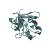
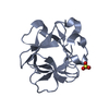
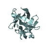
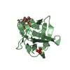
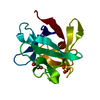
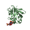
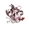
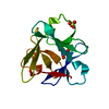
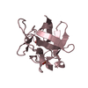
| Title | Crystal Structure Analysis of FGF1-Disaccharide(NI22) complex | |||||||||
 Components Components | Heparin-binding growth factor 1 | |||||||||
 Keywords Keywords |  HORMONE / Heparin/Heparan sulfate Binding HORMONE / Heparin/Heparan sulfate Binding | |||||||||
| Function / homology |  Function and homology information Function and homology informationmesonephric epithelium development / branch elongation involved in ureteric bud branching / regulation of endothelial tube morphogenesis / FGFR3b ligand binding and activation / regulation of endothelial cell chemotaxis to fibroblast growth factor / Signaling by activated point mutants of FGFR3 / FGFR3c ligand binding and activation / Phospholipase C-mediated cascade; FGFR3 / FGFR2b ligand binding and activation / positive regulation of cholesterol biosynthetic process ...mesonephric epithelium development / branch elongation involved in ureteric bud branching / regulation of endothelial tube morphogenesis / FGFR3b ligand binding and activation / regulation of endothelial cell chemotaxis to fibroblast growth factor / Signaling by activated point mutants of FGFR3 / FGFR3c ligand binding and activation / Phospholipase C-mediated cascade; FGFR3 / FGFR2b ligand binding and activation / positive regulation of cholesterol biosynthetic process / fibroblast growth factor receptor binding / FGFR2c ligand binding and activation / Activated point mutants of FGFR2 / Phospholipase C-mediated cascade; FGFR2 / FGFR4 ligand binding and activation / FGFR1b ligand binding and activation / Phospholipase C-mediated cascade; FGFR4 / Signaling by activated point mutants of FGFR1 / FGFR1c ligand binding and activation / organ induction / Downstream signaling of activated FGFR1 / Phospholipase C-mediated cascade: FGFR1 / positive regulation of hepatocyte proliferation / S100 protein binding / positive regulation of intracellular signal transduction / Signaling by FGFR2 IIIa TM / PI-3K cascade:FGFR3 / positive regulation of sprouting angiogenesis / PI-3K cascade:FGFR2 / PI-3K cascade:FGFR4 / PI-3K cascade:FGFR1 / positive regulation of cell division / PI3K Cascade / anatomical structure morphogenesis / fibroblast growth factor receptor signaling pathway / SHC-mediated cascade:FGFR3 / SHC-mediated cascade:FGFR2 / SHC-mediated cascade:FGFR4 / SHC-mediated cascade:FGFR1 / FRS-mediated FGFR3 signaling / FRS-mediated FGFR2 signaling / FRS-mediated FGFR4 signaling / Signaling by FGFR3 in disease / FRS-mediated FGFR1 signaling / Hsp70 protein binding / Signaling by FGFR2 in disease / Signaling by FGFR1 in disease / activation of protein kinase B activity / extracellular matrix / positive regulation of endothelial cell migration / epithelial cell proliferation / positive regulation of epithelial cell proliferation / Negative regulation of FGFR3 signaling / Negative regulation of FGFR2 signaling / Negative regulation of FGFR4 signaling / Negative regulation of FGFR1 signaling / animal organ morphogenesis / lung development / growth factor activity / positive regulation of MAP kinase activity / wound healing / positive regulation of angiogenesis / Constitutive Signaling by Aberrant PI3K in Cancer / integrin binding / PIP3 activates AKT signaling / cellular response to heat / heparin binding / cell cortex / PI5P, PP2A and IER3 Regulate PI3K/AKT Signaling / RAF/MAP kinase cascade / angiogenesis / cell differentiation / positive regulation of ERK1 and ERK2 cascade / positive regulation of cell migration / positive regulation of cell population proliferation / positive regulation of gene expression / signal transduction / positive regulation of transcription by RNA polymerase II / extracellular space / extracellular region / nucleoplasm / nucleus / cytosol / cytoplasmSimilarity search - Function | |||||||||
| Biological species |  Homo sapiens (human) Homo sapiens (human) | |||||||||
| Method | X-RAY DIFFRACTION / SYNCHROTRON / MOLECULAR REPLACEMENT / Resolution: 2.37 Å | |||||||||
 Authors Authors | Hung, S.-C. / Shi, Z. | |||||||||
 Citation Citation | Journal: J.Am.Chem.Soc. / Year: 2012 Title: Divergent synthesis of 48 heparan sulfate-based disaccharides and probing the specific sugar-fibroblast growth factor-1 interaction Authors: Hu, Y.-P. / Zhong, Y.-Q. / Chen, Z.-G. / Chen, C.-Y. / Shi, Z. / Zulueta, M.M.L. / Ku, C.-C. / Lee, P.-Y. / Wang, C.-C. / Hung, S.-C. | |||||||||
| History |
|
- Structure visualization
| Structure viewer | Molecule: MolmilJmol/JSmol |
|---|
- Downloads & links
-Download
| PDBx/mmCIF format | 3ud8.cif.gz | 169.4 KB | Display | PDBx/mmCIF format |
|---|---|---|---|---|
| PDB format | pdb3ud8.ent.gz | 134.5 KB | Display | PDB format |
| PDBx/mmJSON format | 3ud8.json.gz | Tree view | PDBx/mmJSON format | |
| Others |  Other downloads Other downloads |
-Validation report
| Arichive directory | https://data.pdbj.org/pub/pdb/validation_reports/ud/3ud8ftp://data.pdbj.org/pub/pdb/validation_reports/ud/3ud8 | HTTPS FTP |
|---|
-Related structure data
| Related structure data | 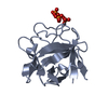 3ud7C 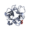 3ud9C  3udaC  1rg8S C: citing same article ( S: Starting model for refinement |
|---|---|
| Similar structure data |

-Links
 PDBj
PDBj


- Assembly
| Deposited unit | 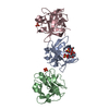
| ||||||||
|---|---|---|---|---|---|---|---|---|---|
| 1 | 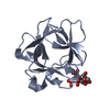
| ||||||||
| 2 | 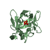
| ||||||||
| 3 | 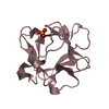
| ||||||||
| Unit cell |
|
-Components
| #1: Protein | Mass: 15989.062 Da / Num. of mol.: 3 Source method: isolated from a genetically manipulated source Source: (gene. exp.) Homo sapiens (human) / Gene: FGF1 / Plasmid: pET21b / Production host:  Escherichia coli (E. coli) / Strain (production host): BL21(DE3) / References: UniProt: P05230 Escherichia coli (E. coli) / Strain (production host): BL21(DE3) / References: UniProt: P05230#2: Polysaccharide | 2-deoxy-3-O-sulfo-2-(sulfoamino)-alpha-D-glucopyranose-(1-4)-1-O-methyl-2-O-sulfo-alpha-L-idopyranuronic acid | / Mass: 609.512 Da / Num. of mol.: 1Source method: isolated from a genetically manipulated source #3: Chemical | Phosphate  Mass: 94.971 Da / Num. of mol.: 2 / Source method: obtained synthetically / Formula: PO4 Mass: 94.971 Da / Num. of mol.: 2 / Source method: obtained synthetically / Formula: PO4#4: Water | ChemComp-HOH / | Water Mass: 18.015 Da / Num. of mol.: 52 / Source method: isolated from a natural source / Formula: H2O Mass: 18.015 Da / Num. of mol.: 52 / Source method: isolated from a natural source / Formula: H2O |
|---|
-Experimental details
-Experiment
| Experiment | Method: X-RAY DIFFRACTION / Number of used crystals: 1 |
|---|
- Sample preparation
| Crystal | Density Matthews: 2.14 Å3/Da / Density % sol: 42.46 % |
|---|---|
| Crystal grow | Temperature: 298 K / Method: vapor diffusion, hanging drop / pH: 7 Details: 2 M (NH4)HPO4, 0.1 M sodium acetate, pH 7.0, VAPOR DIFFUSION, HANGING DROP, temperature 298K |
-Data collection
| Diffraction | Mean temperature: 100 K |
|---|---|
| Diffraction source | Source: SYNCHROTRON / Site: NSRRC  / Beamline: BL13B1 / Wavelength: 1 Å / Beamline: BL13B1 / Wavelength: 1 Å |
| Detector | Type: ADSC QUANTUM 315r / Detector: CCD / Date: Dec 5, 2010 / Details: mirrors |
| Radiation | Monochromator: Si 111 CHANNEL / Protocol: SINGLE WAVELENGTH / Monochromatic (M) / Laue (L): M / Scattering type: x-ray |
| Radiation wavelength | Wavelength: 1 Å / Relative weight: 1 |
| Reflection | Resolution: 2.37→78.43 Å / Num. all: 15943 / Num. obs: 15908 / % possible obs: 95.5 % / Observed criterion σ(F): 0 / Observed criterion σ(I): 5 / Redundancy: 3.4 % / Rmerge(I) obs: 0.055 / Rsym value: 0.055 / Net I/σ(I): 14 |
| Reflection shell | Resolution: 2.38→2.47 Å / Redundancy: 2.7 % / Rmerge(I) obs: 0.241 / Mean I/σ(I) obs: 3.06 / Num. unique all: 1272 / Rsym value: 0.241 / % possible all: 78.2 |
- Processing
| Software |
| |||||||||||||||||||||||||||||||||||||||||||||||||||||||||||||||||||||||||||||||||||||||||||||||||||||||||||||||||||||||||||||||||||||||||||||||||||||||||||||||||||||||||||||||
|---|---|---|---|---|---|---|---|---|---|---|---|---|---|---|---|---|---|---|---|---|---|---|---|---|---|---|---|---|---|---|---|---|---|---|---|---|---|---|---|---|---|---|---|---|---|---|---|---|---|---|---|---|---|---|---|---|---|---|---|---|---|---|---|---|---|---|---|---|---|---|---|---|---|---|---|---|---|---|---|---|---|---|---|---|---|---|---|---|---|---|---|---|---|---|---|---|---|---|---|---|---|---|---|---|---|---|---|---|---|---|---|---|---|---|---|---|---|---|---|---|---|---|---|---|---|---|---|---|---|---|---|---|---|---|---|---|---|---|---|---|---|---|---|---|---|---|---|---|---|---|---|---|---|---|---|---|---|---|---|---|---|---|---|---|---|---|---|---|---|---|---|---|---|---|---|---|
| Refinement | Method to determine structure: MOLECULAR REPLACEMENT Starting model: PDB ENTRY 1RG8 Resolution: 2.37→78.43 Å / Cor.coef. Fo:Fc: 0.944 / Cor.coef. Fo:Fc free: 0.9 / SU B: 20.159 / SU ML: 0.216 / Cross valid method: THROUGHOUT / σ(F): 0 / ESU R Free: 0.318 / Stereochemistry target values: MAXIMUM LIKELIHOOD / Details: HYDROGENS HAVE BEEN ADDED IN THE RIDING POSITIONS
| |||||||||||||||||||||||||||||||||||||||||||||||||||||||||||||||||||||||||||||||||||||||||||||||||||||||||||||||||||||||||||||||||||||||||||||||||||||||||||||||||||||||||||||||
| Solvent computation | Ion probe radii: 0.8 Å / Shrinkage radii: 0.8 Å / VDW probe radii: 1.4 Å / Solvent model: MASK | |||||||||||||||||||||||||||||||||||||||||||||||||||||||||||||||||||||||||||||||||||||||||||||||||||||||||||||||||||||||||||||||||||||||||||||||||||||||||||||||||||||||||||||||
| Displacement parameters | Biso mean: 44.927 Å2
| |||||||||||||||||||||||||||||||||||||||||||||||||||||||||||||||||||||||||||||||||||||||||||||||||||||||||||||||||||||||||||||||||||||||||||||||||||||||||||||||||||||||||||||||
| Refinement step | Cycle: LAST / Resolution: 2.37→78.43 Å
| |||||||||||||||||||||||||||||||||||||||||||||||||||||||||||||||||||||||||||||||||||||||||||||||||||||||||||||||||||||||||||||||||||||||||||||||||||||||||||||||||||||||||||||||
| Refine LS restraints |
| |||||||||||||||||||||||||||||||||||||||||||||||||||||||||||||||||||||||||||||||||||||||||||||||||||||||||||||||||||||||||||||||||||||||||||||||||||||||||||||||||||||||||||||||
| LS refinement shell | Resolution: 2.374→2.436 Å / Total num. of bins used: 20
| |||||||||||||||||||||||||||||||||||||||||||||||||||||||||||||||||||||||||||||||||||||||||||||||||||||||||||||||||||||||||||||||||||||||||||||||||||||||||||||||||||||||||||||||
| Refinement TLS params. | Method: refined / Refine-ID: X-RAY DIFFRACTION
| |||||||||||||||||||||||||||||||||||||||||||||||||||||||||||||||||||||||||||||||||||||||||||||||||||||||||||||||||||||||||||||||||||||||||||||||||||||||||||||||||||||||||||||||
| Refinement TLS group |
|
