DNA topoisomerase / DNA topoisomerase type I (single strand cut, ATP-independent) activity / metabolic process / DNA helicase activity / DNA helicase / ATP hydrolysis activity / DNA binding / ATP binding / metal ion binding / cytoplasm Similarity search - Function
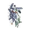
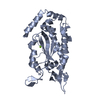
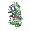
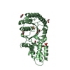
Single alpha-helices involved in coiled-coils or other helix-helix interfaces - #110 / DNA helicase, TraI type / Conjugative transfer relaxase protein TraI / TraI, 2B/2B-like domain / TraI, N-terminal subdomain / DNA helicase TraI, C-terminal / single-stranded DNA binding TraI N-terminal subdomain / DNA relaxase TraI 2B/2B-like domain / Conjugative relaxase, N-terminal / TrwC relaxase ...Single alpha-helices involved in coiled-coils or other helix-helix interfaces - #110 / DNA helicase, TraI type / Conjugative transfer relaxase protein TraI / TraI, 2B/2B-like domain / TraI, N-terminal subdomain / DNA helicase TraI, C-terminal / single-stranded DNA binding TraI N-terminal subdomain / DNA relaxase TraI 2B/2B-like domain / Conjugative relaxase, N-terminal / TrwC relaxase / TrwC relaxase / AAA domain / Single alpha-helices involved in coiled-coils or other helix-helix interfaces / Helix non-globular / Special / P-loop containing nucleoside triphosphate hydrolase Similarity search - Domain/homology
Mass: 18.015 Da / Num. of mol.: 45 / Source method: isolated from a natural source / Formula: H2O
-
Experimental details
-
Experiment
Experiment
Method: X-RAY DIFFRACTION / Number of used crystals: 1
-
Sample preparation
Crystal
Density Matthews: 1.84 Å3/Da / Density % sol: 33.2 %
Crystal grow
Temperature: 298 K / Method: vapor diffusion, hanging drop / pH: 7.5 Details: protein buffer: 50 mM NaCl, 10% glycerol, 10 mM Tris-HCl pH 7.5, mother liquor: 75 mM sodium nitrate, 14% w/v PEG 3350, 10 mM spermine, 110 uM oriT DNA soaked (24 hours): 200 mM ammonium ...Details: protein buffer: 50 mM NaCl, 10% glycerol, 10 mM Tris-HCl pH 7.5, mother liquor: 75 mM sodium nitrate, 14% w/v PEG 3350, 10 mM spermine, 110 uM oriT DNA soaked (24 hours): 200 mM ammonium nitrate, 40% w/v PEG 3350, and 1 mM N,N-imidobisphosphonate (PNP), VAPOR DIFFUSION, HANGING DROP, temperature 298K
In the structure databanks used in Yorodumi, some data are registered as the other names, "COVID-19 virus" and "2019-nCoV". Here are the details of the virus and the list of structure data.
Jan 31, 2019. EMDB accession codes are about to change! (news from PDBe EMDB page)
EMDB accession codes are about to change! (news from PDBe EMDB page)
The allocation of 4 digits for EMDB accession codes will soon come to an end. Whilst these codes will remain in use, new EMDB accession codes will include an additional digit and will expand incrementally as the available range of codes is exhausted. The current 4-digit format prefixed with “EMD-” (i.e. EMD-XXXX) will advance to a 5-digit format (i.e. EMD-XXXXX), and so on. It is currently estimated that the 4-digit codes will be depleted around Spring 2019, at which point the 5-digit format will come into force.
The EM Navigator/Yorodumi systems omit the EMD- prefix.
Related info.:Q: What is EMD? / ID/Accession-code notation in Yorodumi/EM Navigator
Yorodumi is a browser for structure data from EMDB, PDB, SASBDB, etc.
This page is also the successor to EM Navigator detail page, and also detail information page/front-end page for Omokage search.
The word "yorodu" (or yorozu) is an old Japanese word meaning "ten thousand". "mi" (miru) is to see.
Related info.:EMDB / PDB / SASBDB / Comparison of 3 databanks / Yorodumi Search / Aug 31, 2016. New EM Navigator & Yorodumi / Yorodumi Papers / Jmol/JSmol / Function and homology information / Changes in new EM Navigator and Yorodumi
 Movie
Movie Controller

 Yorodumi
 Open data
 Basic information
 Components
 Keywords
 HYDROLASE /
  Function and homology information
 Authors
 Citation
 Structure visualization
 Downloads & links
 Other downloads


 PDBj
PDBj


 Assembly

 Mass: 24.305 Da / Num. of mol.: 1 / Source method: obtained synthetically / Formula: Mg
 Mass: 174.974 Da / Num. of mol.: 1 / Source method: obtained synthetically / Formula: H3NO6P2
 Mass: 322.208 Da / Num. of mol.: 1 / Source method: obtained synthetically / Formula: C10H15N2O8P
 Mass: 18.015 Da / Num. of mol.: 45 / Source method: isolated from a natural source / Formula: H2O
 Sample preparation
 / Beamline: 23-ID-B / Wavelength: 0.97173 Å
 Processing