 Movie
Movie Controller

[English] 日本語
 Yorodumi
- PDB-2q6w: The structure of HLA-DRA, DRB3*0101 (DR52a) with bound platelet i... -
+ Open data

- Basic information
| Entry | Database: PDB / ID: 2q6w | ||||||
|---|---|---|---|---|---|---|---|
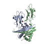
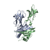
| Title | The structure of HLA-DRA, DRB3*0101 (DR52a) with bound platelet integrin peptide associated with fetal and neonatal alloimmune thrombocytopenia | ||||||
 Components Components |
| ||||||
 Keywords Keywords |  IMMUNE SYSTEM / DRB3 / peptide-class II MHC complex / fetal-maternal alloimmune thrombocytopenia / thrombocytopenia purpura / myasthenia gravis / beta 11 arginine / autoimmunity IMMUNE SYSTEM / DRB3 / peptide-class II MHC complex / fetal-maternal alloimmune thrombocytopenia / thrombocytopenia purpura / myasthenia gravis / beta 11 arginine / autoimmunity | ||||||
| Function / homology |  Function and homology information Function and homology informationtube development / regulation of serotonin uptake / positive regulation of adenylate cyclase-inhibiting opioid receptor signaling pathway / alpha9-beta1 integrin-ADAM8 complex / regulation of trophoblast cell migration / regulation of postsynaptic neurotransmitter receptor diffusion trapping / positive regulation of T cell mediated immune response to tumor cell / alphav-beta3 integrin-vitronectin complex / myeloid dendritic cell antigen processing and presentation / antigen processing and presentation of endogenous peptide antigen via MHC class II ...tube development / regulation of serotonin uptake / positive regulation of adenylate cyclase-inhibiting opioid receptor signaling pathway / alpha9-beta1 integrin-ADAM8 complex / regulation of trophoblast cell migration / regulation of postsynaptic neurotransmitter receptor diffusion trapping / positive regulation of T cell mediated immune response to tumor cell / alphav-beta3 integrin-vitronectin complex / myeloid dendritic cell antigen processing and presentation / antigen processing and presentation of endogenous peptide antigen via MHC class II / autolysosome membrane / maintenance of postsynaptic specialization structure / regulation of T-helper cell differentiation / regulation of extracellular matrix organization / platelet alpha granule membrane / positive regulation of glomerular mesangial cell proliferation / positive regulation of CD4-positive, CD25-positive, alpha-beta regulatory T cell differentiation / integrin alphav-beta3 complex / negative regulation of lipoprotein metabolic process / MHC class II receptor activity / alphav-beta3 integrin-PKCalpha complex / fibrinogen binding / glycinergic synapse / positive regulation of CD4-positive, alpha-beta T cell activation / antigen processing and presentation of peptide or polysaccharide antigen via MHC class II / alphav-beta3 integrin-HMGB1 complex / blood coagulation, fibrin clot formation / negative regulation of lipid transport / vascular endothelial growth factor receptor 2 binding / positive regulation of memory T cell differentiation / negative regulation of low-density lipoprotein receptor activity / regulation of release of sequestered calcium ion into cytosol / angiogenesis involved in wound healing / Elastic fibre formation / cell-substrate junction assembly / mesodermal cell differentiation / alphav-beta3 integrin-IGF-1-IGF1R complex / platelet-derived growth factor receptor binding / filopodium membrane / positive regulation of fibroblast migration / extracellular matrix binding / regulation of postsynaptic neurotransmitter receptor internalization / positive regulation of vascular endothelial growth factor receptor signaling pathway / apolipoprotein A-I-mediated signaling pathway / regulation of bone resorption / apoptotic cell clearance / wound healing, spreading of epidermal cells / positive regulation of cell adhesion mediated by integrin / heterotypic cell-cell adhesion / integrin complex / Molecules associated with elastic fibres / positive regulation of cell-matrix adhesion / cellular response to insulin-like growth factor stimulus / smooth muscle cell migration / transport vesicle membrane / microvillus membrane / cell adhesion mediated by integrin / Syndecan interactions / negative chemotaxis / polysaccharide binding / p130Cas linkage to MAPK signaling for integrins / activation of protein kinase activity / cellular response to platelet-derived growth factor stimulus / cell-substrate adhesion / protein disulfide isomerase activity / positive regulation of smooth muscle cell migration / Translocation of ZAP-70 to Immunological synapse / Phosphorylation of CD3 and TCR zeta chains / positive regulation of osteoblast proliferation / TGF-beta receptor signaling activates SMADs / PECAM1 interactions / lamellipodium membrane / GRB2:SOS provides linkage to MAPK signaling for Integrins / negative regulation of macrophage derived foam cell differentiation / negative regulation of lipid storage / platelet-derived growth factor receptor signaling pathway / fibronectin binding / Generation of second messenger molecules / immunological synapse / ECM proteoglycans / PD-1 signaling / positive regulation of T cell migration / positive regulation of bone resorption / Integrin cell surface interactions / coreceptor activity / negative regulation of endothelial cell apoptotic process / T cell receptor binding / positive regulation of substrate adhesion-dependent cell spreading / cell adhesion molecule binding / positive regulation of endothelial cell proliferation / MHC class II antigen presentation / embryo implantation / positive regulation of endothelial cell migration / Integrin signaling / substrate adhesion-dependent cell spreading / cell-matrix adhesion / trans-Golgi network membrane / response to activity / Signal transduction by L1 / integrin-mediated signaling pathwaySimilarity search - Function | ||||||
| Biological species |  Homo sapiens (human) Homo sapiens (human) | ||||||
| Method | X-RAY DIFFRACTION / SYNCHROTRON / MOLECULAR REPLACEMENT / Resolution: 2.25 Å | ||||||
 Authors Authors | Parry, C.S. | ||||||
 Citation Citation | Journal: J.Mol.Biol. / Year: 2007 Title: Crystallographic Structure of the Human Leukocyte Antigen DRA, DRB3*0101: Models of a Directional Alloimmune Response and Autoimmunity Authors: Parry, C.S. / Gorski, J. / Stern, L.J. | ||||||
| History |
|
- Structure visualization
| Structure viewer | Molecule: MolmilJmol/JSmol |
|---|
- Downloads & links
-Download
| PDBx/mmCIF format | 2q6w.cif.gz | 174 KB | Display | PDBx/mmCIF format |
|---|---|---|---|---|
| PDB format | pdb2q6w.ent.gz | 137.3 KB | Display | PDB format |
| PDBx/mmJSON format | 2q6w.json.gz | Tree view | PDBx/mmJSON format | |
| Others |  Other downloads Other downloads |
-Validation report
| Arichive directory | https://data.pdbj.org/pub/pdb/validation_reports/q6/2q6wftp://data.pdbj.org/pub/pdb/validation_reports/q6/2q6w | HTTPS FTP |
|---|
-Related structure data
| Related structure data |  1dlhS S: Starting model for refinement |
|---|---|
| Similar structure data |

-Links
 PDBj
PDBj


- Assembly
| Deposited unit | 
| ||||||||
|---|---|---|---|---|---|---|---|---|---|
| 1 | 
| ||||||||
| 2 | 
| ||||||||
| Unit cell |
|
-Components
| #1: Protein | Mass: 21155.904 Da / Num. of mol.: 2 / Fragment: sequence database residues 26-207 Source method: isolated from a genetically manipulated source Source: (gene. exp.) Homo sapiens (human) / Gene: HLA-DRA / Production host:   Spodoptera frugiperda (fall armyworm) / Strain (production host): S2 / References: UniProt: P01903 Spodoptera frugiperda (fall armyworm) / Strain (production host): S2 / References: UniProt: P01903#2: Protein | Mass: 22102.637 Da / Num. of mol.: 2 / Fragment: sequence database residues 30-219 Source method: isolated from a genetically manipulated source Source: (gene. exp.) Homo sapiens (human) / Gene: HLA-DRB3 / Production host: Spodoptera frugiperda (fall armyworm) / Strain (production host): S2 / References: UniProt: P79483#3: Protein/peptide | Integrin beta 3 / Platelet membrane glycoprotein IIIa / GPIIIa / CD61 antigenMass: 1302.414 Da / Num. of mol.: 2 / Fragment: sequence database residues 50-61 / Mutation: C26R / Source method: obtained synthetically / Source: (synth.) Homo sapiens (human) / References: UniProt: P05106#4: Water | ChemComp-HOH / | Water Mass: 18.015 Da / Num. of mol.: 487 / Source method: isolated from a natural source / Formula: H2O Mass: 18.015 Da / Num. of mol.: 487 / Source method: isolated from a natural source / Formula: H2O |
|---|
-Experimental details
-Experiment
| Experiment | Method: X-RAY DIFFRACTION / Number of used crystals: 1 |
|---|
- Sample preparation
| Crystal | Density Matthews: 2.96 Å3/Da / Density % sol: 58.45 % |
|---|---|
| Crystal grow | Temperature: 297 K / Method: vapor diffusion, hanging drop / pH: 4.4 Details: 10-20% PEG 8000, 0.1M sodium acetate, 10% (v/v) glycerol, 0.01M Cd2+, pH 4.4, VAPOR DIFFUSION, HANGING DROP, temperature 297.0K |
-Data collection
| Diffraction | Mean temperature: 98 K |
|---|---|
| Diffraction source | Source: SYNCHROTRON / Site: NSLS  / Beamline: X25 / Wavelength: 1.1 Å / Beamline: X25 / Wavelength: 1.1 Å |
| Detector | Type: ADSC QUANTUM 4 / Detector: CCD / Date: Jan 31, 2002 |
| Radiation | Protocol: SINGLE WAVELENGTH / Monochromatic (M) / Laue (L): M / Scattering type: x-ray |
| Radiation wavelength | Wavelength: 1.1 Å / Relative weight: 1 |
| Reflection | Resolution: 2.25→30 Å / Num. all: 51116 / Num. obs: 48049 / % possible obs: 94 % / Observed criterion σ(F): 2 / Observed criterion σ(I): 2 / Redundancy: 15.8 % / Biso Wilson estimate: 42.9 Å2 / Rmerge(I) obs: 0.12 |
| Reflection shell | Resolution: 2.25→2.308 Å / Rmerge(I) obs: 0.36 / Mean I/σ(I) obs: 3.04 / % possible all: 55.7 |
- Processing
| Software |
| ||||||||||||||||||||||||||||||||||||||||||||||||||||||||||||||||||||||||||||||||||||||||||
|---|---|---|---|---|---|---|---|---|---|---|---|---|---|---|---|---|---|---|---|---|---|---|---|---|---|---|---|---|---|---|---|---|---|---|---|---|---|---|---|---|---|---|---|---|---|---|---|---|---|---|---|---|---|---|---|---|---|---|---|---|---|---|---|---|---|---|---|---|---|---|---|---|---|---|---|---|---|---|---|---|---|---|---|---|---|---|---|---|---|---|---|
| Refinement | Method to determine structure: MOLECULAR REPLACEMENT Starting model: PDB entry 1DLH Resolution: 2.25→20.05 Å / Cor.coef. Fo:Fc: 0.943 / Cor.coef. Fo:Fc free: 0.912 / SU B: 5.91 / SU ML: 0.15 / Cross valid method: THROUGHOUT / σ(F): 2 / ESU R: 0.295 / ESU R Free: 0.24 / Stereochemistry target values: MAXIMUM LIKELIHOOD / Details: HYDROGENS HAVE BEEN ADDED IN THE RIDING POSITIONS
| ||||||||||||||||||||||||||||||||||||||||||||||||||||||||||||||||||||||||||||||||||||||||||
| Solvent computation | Ion probe radii: 0.8 Å / Shrinkage radii: 0.8 Å / VDW probe radii: 1.2 Å / Solvent model: MASK | ||||||||||||||||||||||||||||||||||||||||||||||||||||||||||||||||||||||||||||||||||||||||||
| Displacement parameters | Biso mean: 42.241 Å2
| ||||||||||||||||||||||||||||||||||||||||||||||||||||||||||||||||||||||||||||||||||||||||||
| Refinement step | Cycle: LAST / Resolution: 2.25→20.05 Å
| ||||||||||||||||||||||||||||||||||||||||||||||||||||||||||||||||||||||||||||||||||||||||||
| Refine LS restraints |
| ||||||||||||||||||||||||||||||||||||||||||||||||||||||||||||||||||||||||||||||||||||||||||
| LS refinement shell | Resolution: 2.25→2.308 Å / Total num. of bins used: 20
|
