 Movie
Movie Controller

[English] 日本語
 Yorodumi
- PDB-2pyq: Crystal structure of a duf2853 member protein (jann_4075) from ja... -
+ Open data

- Basic information
| Entry | Database: PDB / ID: 2pyq | ||||||
|---|---|---|---|---|---|---|---|
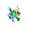
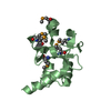
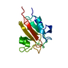
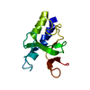
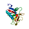
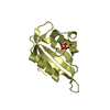
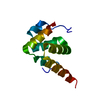
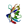
| Title | Crystal structure of a duf2853 member protein (jann_4075) from jannaschia sp. ccs1 at 1.500 A resolution | ||||||
 Components Components | Uncharacterized protein | ||||||
 Keywords Keywords | UNKNOWN FUNCTION /  Structural genomics / Joint Center for Structural Genomics / JCSG / Protein Structure Initiative / PSI-2 Structural genomics / Joint Center for Structural Genomics / JCSG / Protein Structure Initiative / PSI-2 | ||||||
| Function / homology | Jann4075-like / Protein of unknown function DUF2853 / Jann4075-like superfamily / Protein of unknown function (DUF2853) / Recoverin; domain 1 / Orthogonal Bundle / Mainly Alpha / Uncharacterized protein Function and homology information Function and homology information | ||||||
| Biological species |  Jannaschia sp. CCS1 (bacteria) Jannaschia sp. CCS1 (bacteria) | ||||||
| Method | X-RAY DIFFRACTION / SYNCHROTRON / MAD / Resolution: 1.5 Å | ||||||
 Authors Authors | Joint Center for Structural Genomics (JCSG) | ||||||
 Citation Citation | Journal: To be published Title: Crystal structure of uncharacterized protein (YP_512017.1) from Jannaschia sp. CCS1 at 1.500 A resolution Authors: Joint Center for Structural Genomics (JCSG) | ||||||
| History |
| ||||||
| Remark 999 | SEQUENCE THE CONSTRUCT WAS EXPRESSED WITH A PURIFICATION TAG MGSDKIHHHHHHENLYFQG. THE TAG WAS ... SEQUENCE THE CONSTRUCT WAS EXPRESSED WITH A PURIFICATION TAG MGSDKIHHHHHHENLYFQG. THE TAG WAS REMOVED WITH TEV PROTEASE LEAVING ONLY A GLYCINE (0) FOLLOWED BY THE TARGET SEQUENCE. |
- Structure visualization
| Structure viewer | Molecule: MolmilJmol/JSmol |
|---|
- Downloads & links
-Download
| PDBx/mmCIF format | 2pyq.cif.gz | 186.4 KB | Display | PDBx/mmCIF format |
|---|---|---|---|---|
| PDB format | pdb2pyq.ent.gz | 154 KB | Display | PDB format |
| PDBx/mmJSON format | 2pyq.json.gz | Tree view | PDBx/mmJSON format | |
| Others |  Other downloads Other downloads |
-Validation report
| Arichive directory | https://data.pdbj.org/pub/pdb/validation_reports/py/2pyqftp://data.pdbj.org/pub/pdb/validation_reports/py/2pyq | HTTPS FTP |
|---|
-Related structure data
| Similar structure data | |
|---|---|
| Other databases |

-Links
 PDBj
PDBj- Assembly
| Deposited unit | 
| ||||||||
|---|---|---|---|---|---|---|---|---|---|
| 1 | 
| ||||||||
| 2 | 
| ||||||||
| 3 | 
| ||||||||
| 4 | 
| ||||||||
| Unit cell |
|
-Components
| #1: Protein | Mass: 12924.854 Da / Num. of mol.: 4 Source method: isolated from a genetically manipulated source Source: (gene. exp.) Jannaschia sp. CCS1 (bacteria) / Gene: YP_512017.1, Jann_4075 / Plasmid: speedET / Production host: Escherichia coli (E. coli) / Strain (production host): HK100 / References: UniProt: Q28JX0#2: Chemical | Polyethylene glycol  Mass: 194.226 Da / Num. of mol.: 2 / Source method: obtained synthetically / Formula: C8H18O5 / Comment: precipitant*YM Mass: 194.226 Da / Num. of mol.: 2 / Source method: obtained synthetically / Formula: C8H18O5 / Comment: precipitant*YM#3: Water | ChemComp-HOH / | Water Mass: 18.015 Da / Num. of mol.: 304 / Source method: isolated from a natural source / Formula: H2O Mass: 18.015 Da / Num. of mol.: 304 / Source method: isolated from a natural source / Formula: H2O |
|---|
-Experimental details
-Experiment
| Experiment | Method: X-RAY DIFFRACTION / Number of used crystals: 1 |
|---|
- Sample preparation
| Crystal | Density Matthews: 1.84 Å3/Da / Density % sol: 33.15 % |
|---|---|
| Crystal grow | Temperature: 277 K / Method: vapor diffusion, sitting drop / pH: 7 Details: NANODROP, 30.0% PEG 6000, 0.1M HEPES pH 7.0, VAPOR DIFFUSION, SITTING DROP, temperature 277K |
-Data collection
| Diffraction | Mean temperature: 100 K | |||||||||||||||||||||||||||||||||||||||||||||||||||||||||||||||||||||||||||||
|---|---|---|---|---|---|---|---|---|---|---|---|---|---|---|---|---|---|---|---|---|---|---|---|---|---|---|---|---|---|---|---|---|---|---|---|---|---|---|---|---|---|---|---|---|---|---|---|---|---|---|---|---|---|---|---|---|---|---|---|---|---|---|---|---|---|---|---|---|---|---|---|---|---|---|---|---|---|---|
| Diffraction source | Source: SYNCHROTRON / Site: SSRL  / Beamline: BL11-1 / Wavelength: 0.91837, 0.97910, 0.97886 / Beamline: BL11-1 / Wavelength: 0.91837, 0.97910, 0.97886 | |||||||||||||||||||||||||||||||||||||||||||||||||||||||||||||||||||||||||||||
| Detector | Type: MARMOSAIC 325 mm CCD / Detector: CCD / Date: Apr 8, 2007 / Details: Flat mirror (vertical focusing) | |||||||||||||||||||||||||||||||||||||||||||||||||||||||||||||||||||||||||||||
| Radiation | Monochromator: Single crystal Si(111) bent (horizontal focusing) Protocol: MAD / Monochromatic (M) / Laue (L): M / Scattering type: x-ray | |||||||||||||||||||||||||||||||||||||||||||||||||||||||||||||||||||||||||||||
| Radiation wavelength |
| |||||||||||||||||||||||||||||||||||||||||||||||||||||||||||||||||||||||||||||
| Reflection | Resolution: 1.5→28.689 Å / Num. obs: 59568 / % possible obs: 97.4 % / Observed criterion σ(I): -3 / Biso Wilson estimate: 28.524 Å2 / Rmerge(I) obs: 0.052 / Net I/σ(I): 7.77 | |||||||||||||||||||||||||||||||||||||||||||||||||||||||||||||||||||||||||||||
| Reflection shell |
|
-Phasing
| Phasing | Method: MAD |
|---|
- Processing
| Software |
| |||||||||||||||||||||||||||||||||||||||||||||||||||||||||||||||||||||||||||||||||||||||||||||||||||||||||||||||||||||||||||||
|---|---|---|---|---|---|---|---|---|---|---|---|---|---|---|---|---|---|---|---|---|---|---|---|---|---|---|---|---|---|---|---|---|---|---|---|---|---|---|---|---|---|---|---|---|---|---|---|---|---|---|---|---|---|---|---|---|---|---|---|---|---|---|---|---|---|---|---|---|---|---|---|---|---|---|---|---|---|---|---|---|---|---|---|---|---|---|---|---|---|---|---|---|---|---|---|---|---|---|---|---|---|---|---|---|---|---|---|---|---|---|---|---|---|---|---|---|---|---|---|---|---|---|---|---|---|---|
| Refinement | Method to determine structure: MAD / Resolution: 1.5→28.689 Å / FOM work R set: 0.75 / Stereochemistry target values: twin_lsq_f Details: 1. HYDROGENS HAVE BEEN ADDED IN THE RIDING POSITIONS. 2. A MET-INHIBITION PROTOCOL WAS USED FOR SELENOMETHIONINE INCORPORATION DURING PROTEIN EXPRESSION. THE OCCUPANCY OF THE SE ATOMS IN THE ...Details: 1. HYDROGENS HAVE BEEN ADDED IN THE RIDING POSITIONS. 2. A MET-INHIBITION PROTOCOL WAS USED FOR SELENOMETHIONINE INCORPORATION DURING PROTEIN EXPRESSION. THE OCCUPANCY OF THE SE ATOMS IN THE MSE RESIDUES WAS REDUCED TO 0.75 FOR THE REDUCED SCATTERING POWER DUE TO PARTIAL S-MET INCORPORATION. 3. PEG 200 MOLECULES (PG4) ARE MODELED. 4. THE DATA ARE TWINNED WITH THE TWIN LAW -H,-K,L. THE REFINED TWIN FRACTION WAS 0.227. 5. DUE TO 4-FOLD NCS AND TWINNING, REFLECTIONS FOR R-FREE SET WERE SELECTED IN THIN SHELLS. 6. WHEN REFINING TLS, THE OUTPUT PDB FILE ALWAYS HAS THE ANISOU RECORDS FOR THE ATOMS INVOLVED IN TLS GROUPS. THE ANISOTROPIC B-FACTOR IN ANISOU RECORDS IS THE TOTAL B-FACTOR (B_TLS + B_INDIVIDUAL). THE ISOTROPIC EQUIVALENT B-FACTOR IN ATOM RECORDS IS THE MEAN OF THE TRACE OF THE ANISOU MATRIX DIVIDED BY 10000 AND MULTIPLIED BY 8*PI^2 AND REPRESENTS THE ISOTROPIC EQUIVALENT OF THE TOTAL B-FACTOR (B_TLS + B_INDIVIDUAL). TO OBTAIN THE INDIVIDUAL B-FACTORS, ONE NEEDS TO COMPUTE THE TLS COMPONENT (B_TLS) USING THE TLS RECORDS IN THE PDB FILE HEADER AND THEN SUBTRACT IT FROM THE TOTAL B-FACTORS (ON THE ANISOU RECORDS).
| |||||||||||||||||||||||||||||||||||||||||||||||||||||||||||||||||||||||||||||||||||||||||||||||||||||||||||||||||||||||||||||
| Solvent computation | Bsol: 51.298 Å2 / ksol: 0.396 e/Å3 | |||||||||||||||||||||||||||||||||||||||||||||||||||||||||||||||||||||||||||||||||||||||||||||||||||||||||||||||||||||||||||||
| Displacement parameters | Biso max: 103.1 Å2 / Biso mean: 27.35 Å2 / Biso min: 13.17 Å2
| |||||||||||||||||||||||||||||||||||||||||||||||||||||||||||||||||||||||||||||||||||||||||||||||||||||||||||||||||||||||||||||
| Refinement step | Cycle: LAST / Resolution: 1.5→28.689 Å
| |||||||||||||||||||||||||||||||||||||||||||||||||||||||||||||||||||||||||||||||||||||||||||||||||||||||||||||||||||||||||||||
| Refine LS restraints |
| |||||||||||||||||||||||||||||||||||||||||||||||||||||||||||||||||||||||||||||||||||||||||||||||||||||||||||||||||||||||||||||
| Refinement TLS params. | Method: refined / Refine-ID: X-RAY DIFFRACTION
| |||||||||||||||||||||||||||||||||||||||||||||||||||||||||||||||||||||||||||||||||||||||||||||||||||||||||||||||||||||||||||||
| Refinement TLS group |
|
