 Movie
Movie Controller

[English] 日本語
 Yorodumi
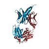
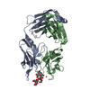
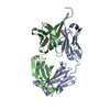
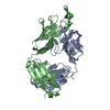
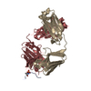
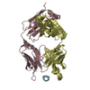
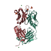
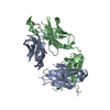
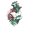
- PDB-2pw1: Crystal structure of the HIV-1 Cross Neutralizing Monoclonal Anti... -
+ Open data

- Basic information
| Entry | Database: PDB / ID: 2pw1 | ||||||
|---|---|---|---|---|---|---|---|
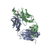
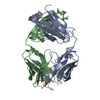
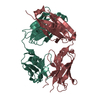
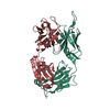
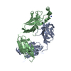
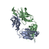
| Title | Crystal structure of the HIV-1 Cross Neutralizing Monoclonal Antibody 2F5 in complex with gp41 Peptide ELDKWNSL | ||||||
 Components Components |
| ||||||
 Keywords Keywords |  IMMUNE SYSTEM / HIV-1 / 2F5 / gp41 / neutralizing antibody IMMUNE SYSTEM / HIV-1 / 2F5 / gp41 / neutralizing antibody | ||||||
| Function / homology | Immunoglobulins / Immunoglobulin-like / Sandwich / Mainly Beta Function and homology information Function and homology information | ||||||
| Biological species |  Homo sapiens (human) Homo sapiens (human) | ||||||
| Method | X-RAY DIFFRACTION / MOLECULAR REPLACEMENT / Resolution: 2.6 Å | ||||||
 Authors Authors | Bryson, S. / Julien, J.-P. / Hynes, R.C. / Pai, E.F. | ||||||
 Citation Citation | Journal: J.Virol. / Year: 2009 Title: Crystallographic definition of the epitope promiscuity of the broadly neutralizing anti-human immunodeficiency virus type 1 antibody 2F5: vaccine design implications. Authors: Bryson, S. / Julien, J.P. / Hynes, R.C. / Pai, E.F. | ||||||
| History |
|
- Structure visualization
| Structure viewer | Molecule: MolmilJmol/JSmol |
|---|
- Downloads & links
-Download
| PDBx/mmCIF format | 2pw1.cif.gz | 96.9 KB | Display | PDBx/mmCIF format |
|---|---|---|---|---|
| PDB format | pdb2pw1.ent.gz | 77.9 KB | Display | PDB format |
| PDBx/mmJSON format | 2pw1.json.gz | Tree view | PDBx/mmJSON format | |
| Others |  Other downloads Other downloads |
-Validation report
| Arichive directory | https://data.pdbj.org/pub/pdb/validation_reports/pw/2pw1ftp://data.pdbj.org/pub/pdb/validation_reports/pw/2pw1 | HTTPS FTP |
|---|
-Related structure data
| Related structure data |  1u8hC  1u8iC  1u8jC  1u8lC  1u8mC  1u8nC 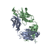 1u8oC  1u8pC  1u8qC  1u91C  1u92C 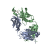 1u93C  1u95C  2f5aC 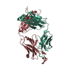 2f5bC 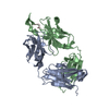 2pw2C  3idgC 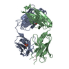 3idiC 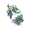 3idjC 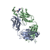 3idmC 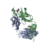 3idnC C: citing same article ( |
|---|---|
| Similar structure data |


-Links
 PDBj
PDBj


- Assembly
| Deposited unit | 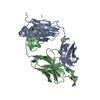
| ||||||||
|---|---|---|---|---|---|---|---|---|---|
| 1 |
| ||||||||
| Unit cell |
| ||||||||
| Details | The heavy chain and the light chain of the antibody assemble to form the 2F5 Fab fragment. the biological unit is identical to the asymmetric unit. |
-Components
| #1: Antibody | Mass: 23363.844 Da / Num. of mol.: 1 / Source method: isolated from a natural source / Source: (natural) Homo sapiens (human) |
|---|---|
| #2: Antibody | Mass: 24985.436 Da / Num. of mol.: 1 / Source method: isolated from a natural source / Source: (natural) Homo sapiens (human) |
| #3: Protein/peptide | Mass: 1005.103 Da / Num. of mol.: 1 / Source method: obtained synthetically / Details: synthetic peptide |
| #4: Water | ChemComp-HOH / Water Mass: 18.015 Da / Num. of mol.: 174 / Source method: isolated from a natural source / Formula: H2O Mass: 18.015 Da / Num. of mol.: 174 / Source method: isolated from a natural source / Formula: H2O |
-Experimental details
-Experiment
| Experiment | Method: X-RAY DIFFRACTION / Number of used crystals: 1 |
|---|
- Sample preparation
| Crystal | Density Matthews: 3.36 Å3/Da / Density % sol: 63.39 % |
|---|---|
| Crystal grow | Temperature: 293 K / Method: vapor diffusion, hanging drop / pH: 5.6 Details: 0.1M NaCitrate, AS 1.6M, pH 5.6, VAPOR DIFFUSION, HANGING DROP, temperature 293K |
-Data collection
| Diffraction | Mean temperature: 100 K |
|---|---|
| Diffraction source | Source: ROTATING ANODE / Type: RIGAKU RUH3R / Wavelength: 1.54 Å |
| Detector | Type: RIGAKU RAXIS IV / Detector: IMAGE PLATE / Date: Dec 10, 2005 |
| Radiation | Protocol: SINGLE WAVELENGTH / Monochromatic (M) / Laue (L): M / Scattering type: x-ray |
| Radiation wavelength | Wavelength: 1.54 Å / Relative weight: 1 |
| Reflection | Resolution: 2.6→45 Å / Num. all: 21089 / Num. obs: 20317 / % possible obs: 96.3 % / Redundancy: 4.5 % / Biso Wilson estimate: 33.2 Å2 / Rsym value: 0.135 |
| Reflection shell | Resolution: 2.6→2.76 Å / Mean I/σ(I) obs: 1.2 / Num. unique all: 2972 / Rsym value: 0.352 |
- Processing
| Software |
| ||||||||||||||||||||||||||||||||||||
|---|---|---|---|---|---|---|---|---|---|---|---|---|---|---|---|---|---|---|---|---|---|---|---|---|---|---|---|---|---|---|---|---|---|---|---|---|---|
| Refinement | Method to determine structure: MOLECULAR REPLACEMENT / Resolution: 2.6→43.78 Å / Rfactor Rfree error: 0.007 / Data cutoff high absF: 1201898.24 / Data cutoff low absF: 0 / Isotropic thermal model: RESTRAINED / Cross valid method: THROUGHOUT / σ(F): 0
| ||||||||||||||||||||||||||||||||||||
| Solvent computation | Solvent model: FLAT MODEL / Bsol: 29.9517 Å2 / ksol: 0.370182 e/Å3 | ||||||||||||||||||||||||||||||||||||
| Displacement parameters | Biso mean: 25.8 Å2
| ||||||||||||||||||||||||||||||||||||
| Refine analyze |
| ||||||||||||||||||||||||||||||||||||
| Refinement step | Cycle: LAST / Resolution: 2.6→43.78 Å
| ||||||||||||||||||||||||||||||||||||
| Refine LS restraints |
| ||||||||||||||||||||||||||||||||||||
| LS refinement shell | Resolution: 2.6→2.76 Å / Rfactor Rfree error: 0.028 / Total num. of bins used: 6
| ||||||||||||||||||||||||||||||||||||
| Xplor file |
|
