 Movie
Movie Controller

[English] 日本語
 Yorodumi
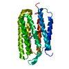
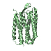
- PDB-2psm: Crystal structure of Interleukin 15 in complex with Interleukin 1... -
+ Open data

- Basic information
| Entry | Database: PDB / ID: 2psm | |||||||||
|---|---|---|---|---|---|---|---|---|---|---|
| Title | Crystal structure of Interleukin 15 in complex with Interleukin 15 receptor alpha | |||||||||
 Components Components |
| |||||||||
 Keywords Keywords |  CYTOKINE / Glycoprotein / Secreted / Alternative splicing / Endoplasmic reticulum / Golgi apparatus / Membrane / Nucleus / Phosphorylation / Receptor / Sushi / Transmembrane / Structural Genomics / NPPSFA / National Project on Protein Structural and Functional Analyses / RIKEN Structural Genomics/Proteomics Initiative / RSGI CYTOKINE / Glycoprotein / Secreted / Alternative splicing / Endoplasmic reticulum / Golgi apparatus / Membrane / Nucleus / Phosphorylation / Receptor / Sushi / Transmembrane / Structural Genomics / NPPSFA / National Project on Protein Structural and Functional Analyses / RIKEN Structural Genomics/Proteomics Initiative / RSGI | |||||||||
| Function / homology |  Function and homology information Function and homology informationNK T cell proliferation / extrathymic T cell selection / positive regulation of protein O-linked glycosylation / hyaluronan metabolic process / Interleukin-15 signaling / positive regulation of natural killer cell differentiation / interleukin-15 receptor activity / skeletal muscle atrophy / : / neutrophil activation ...NK T cell proliferation / extrathymic T cell selection / positive regulation of protein O-linked glycosylation / hyaluronan metabolic process / Interleukin-15 signaling / positive regulation of natural killer cell differentiation / interleukin-15 receptor activity / skeletal muscle atrophy / : / neutrophil activation / cytokine receptor binding / cellular response to vitamin D / regulation of defense response to virus by host / interleukin-15-mediated signaling pathway / tyrosine phosphorylation of STAT protein / negative regulation of cold-induced thermogenesis / regulation of T cell differentiation / positive regulation of natural killer cell proliferation / positive regulation of interleukin-17 production / cell surface receptor signaling pathway via JAK-STAT / macrophage differentiation / lymph node development / positive regulation of phagocytosis / positive regulation of T cell proliferation / cell maturation / positive regulation of tyrosine phosphorylation of STAT protein / cytokine activity / positive regulation of cytokine production / negative regulation of smooth muscle cell proliferation / positive regulation of immune response / positive regulation of peptidyl-tyrosine phosphorylation / negative regulation of neuron projection development / cytoplasmic vesicle / nuclear membrane / membrane => GO:0016020 / nuclear speck / immune response / inflammatory response / positive regulation of cell population proliferation / protein kinase binding / cell surface / extracellular space / extracellular region / nucleoplasm / plasma membrane / cytosol / cytoplasmSimilarity search - Function | |||||||||
| Biological species |  Mus musculus (house mouse) Mus musculus (house mouse) | |||||||||
| Method | X-RAY DIFFRACTION / SYNCHROTRON / SAD / Resolution: 2.19 Å | |||||||||
 Authors Authors | Olsen, S.K. / Murayama, K. / Kishishita, S. / Kukimoto-Niino, M. / Terada, T. / Shirouzu, M. / Ota, N. / Kanagawa, O. / Yokoyama, S. / RIKEN Structural Genomics/Proteomics Initiative (RSGI) | |||||||||
 Citation Citation | Journal: J.Biol.Chem. / Year: 2007 Title: Crystal Structure of the Interleukin-15{middle dot}Interleukin-15 Receptor {alpha} Complex: INSIGHTS INTO TRANS AND CIS PRESENTATION Authors: Olsen, S.K. / Ota, N. / Kishishita, S. / Kukimoto-Niino, M. / Murayama, K. / Uchiyama, H. / Toyama, M. / Terada, T. / Shirouzu, M. / Kanagawa, O. / Yokoyama, S. | |||||||||
| History |
| |||||||||
| Remark 300 | BIOMOLECULE: 1 SEE REMARK 350 FOR THE AUTHOR PROVIDED AND/OR PROGRAM GENERATED ASSEMBLY INFORMATION ...BIOMOLECULE: 1 SEE REMARK 350 FOR THE AUTHOR PROVIDED AND/OR PROGRAM GENERATED ASSEMBLY INFORMATION FOR THE STRUCTURE IN THIS ENTRY. THE REMARK MAY ALSO PROVIDE INFORMATION ON BURIED SURFACE AREA. DETAILS: AUTHOR DETERMINED BIOLOGICAL UNIT: UNKNOWN |
- Structure visualization
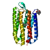
| Structure viewer | Molecule: MolmilJmol/JSmol |
|---|
- Downloads & links
-Download
| PDBx/mmCIF format | 2psm.cif.gz | 82.7 KB | Display | PDBx/mmCIF format |
|---|---|---|---|---|
| PDB format | pdb2psm.ent.gz | 65.6 KB | Display | PDB format |
| PDBx/mmJSON format | 2psm.json.gz | Tree view | PDBx/mmJSON format | |
| Others |  Other downloads Other downloads |
-Validation report
| Arichive directory | https://data.pdbj.org/pub/pdb/validation_reports/ps/2psmftp://data.pdbj.org/pub/pdb/validation_reports/ps/2psm | HTTPS FTP |
|---|
-Related structure data
| Similar structure data | |
|---|---|
| Other databases |


-Links
 PDBj
PDBj


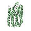
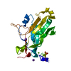
- Assembly
| Deposited unit | 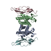
| ||||||||
|---|---|---|---|---|---|---|---|---|---|
| 1 | 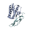
| ||||||||
| 2 | 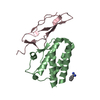
| ||||||||
| Unit cell |
|
-Components
| #1: Protein | Interleukin 15 / IL-15 Mass: 13781.437 Da / Num. of mol.: 2 Source method: isolated from a genetically manipulated source Source: (gene. exp.) Mus musculus (house mouse) / Description: Cell Free Expression: E.coli / Gene: Il15 / Production host: Cell free synthesis / References: UniProt: P48346#2: Protein | Mass: 8378.438 Da / Num. of mol.: 2 / Fragment: UNP residues 33-103 Source method: isolated from a genetically manipulated source Source: (gene. exp.) Mus musculus (house mouse) / Description: Cell Free Expression: E.coli / Gene: Il15ra / Production host: Cell free synthesis / References: UniProt: Q60819#3: Chemical | ChemComp-BEN / | Benzamidine  Mass: 120.152 Da / Num. of mol.: 1 / Source method: obtained synthetically / Formula: C7H8N2 Mass: 120.152 Da / Num. of mol.: 1 / Source method: obtained synthetically / Formula: C7H8N2#4: Water | ChemComp-HOH / | Water Mass: 18.015 Da / Num. of mol.: 91 / Source method: isolated from a natural source / Formula: H2O Mass: 18.015 Da / Num. of mol.: 91 / Source method: isolated from a natural source / Formula: H2O |
|---|
-Experimental details
-Experiment
| Experiment | Method: X-RAY DIFFRACTION / Number of used crystals: 1 |
|---|
- Sample preparation
| Crystal | Density Matthews: 3.01 Å3/Da / Density % sol: 59.16 % Description: THE STRUCTURE FACTOR FILE CONTAINS FRIEDEL PAIRS. |
|---|---|
| Crystal grow | Temperature: 293 K / Method: vapor diffusion, hanging drop / pH: 6.8 Details: 1.7M Na/K Phosphate, pH 6.8, VAPOR DIFFUSION, HANGING DROP, temperature 293K |
-Data collection
| Diffraction | Mean temperature: 100 K |
|---|---|
| Diffraction source | Source: SYNCHROTRON / Site: Photon Factory  / Beamline: AR-NW12A / Wavelength: 0.9793 Å / Beamline: AR-NW12A / Wavelength: 0.9793 Å |
| Detector | Type: ADSC QUANTUM 210 / Detector: CCD / Date: Feb 10, 2007 |
| Radiation | Monochromator: Si (111) double crystal / Protocol: SINGLE WAVELENGTH / Monochromatic (M) / Laue (L): M / Scattering type: x-ray |
| Radiation wavelength | Wavelength: 0.9793 Å / Relative weight: 1 |
| Reflection | Resolution: 2.19→50 Å / Num. obs: 53064 / % possible obs: 99.9 % / Observed criterion σ(I): -3 / Redundancy: 14.4 % / Rsym value: 0.057 / Net I/σ(I): 23 |
| Reflection shell | Resolution: 2.19→2.27 Å / Mean I/σ(I) obs: 18.6 / Num. unique all: 2777 / Rsym value: 0.163 / % possible all: 100 |
- Processing
| Software |
| ||||||||||||||||||||||||||||||||||||||||||||||||||||||||||||
|---|---|---|---|---|---|---|---|---|---|---|---|---|---|---|---|---|---|---|---|---|---|---|---|---|---|---|---|---|---|---|---|---|---|---|---|---|---|---|---|---|---|---|---|---|---|---|---|---|---|---|---|---|---|---|---|---|---|---|---|---|---|
| Refinement | Method to determine structure: SAD / Resolution: 2.19→25 Å / Isotropic thermal model: restrained / Cross valid method: THROUGHOUT / σ(F): 0 / Stereochemistry target values: Engh & Huber / Details: THE STRUCTURE FACTOR FILE CONTAINS FRIEDEL PAIRS.
| ||||||||||||||||||||||||||||||||||||||||||||||||||||||||||||
| Displacement parameters |
| ||||||||||||||||||||||||||||||||||||||||||||||||||||||||||||
| Refinement step | Cycle: LAST / Resolution: 2.19→25 Å
| ||||||||||||||||||||||||||||||||||||||||||||||||||||||||||||
| Refine LS restraints |
|
