 Movie
Movie Controller

[English] 日本語
 Yorodumi
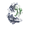
- PDB-2pcj: Crystal structure of ABC transporter (aq_297) From Aquifex Aeolic... -
+ Open data

- Basic information
| Entry | Database: PDB / ID: 2pcj | ||||||
|---|---|---|---|---|---|---|---|
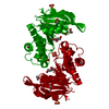
| Title | Crystal structure of ABC transporter (aq_297) From Aquifex Aeolicus VF5 | ||||||
 Components Components | Lipoprotein-releasing system ATP-binding protein lolD | ||||||
 Keywords Keywords |  HYDROLASE / ABC TRANSPORTER / ATP-BINDING PROTEIN / Structural Genomics / NPPSFA / National Project on Protein Structural and Functional Analyses / RIKEN Structural Genomics/Proteomics Initiative / RSGI HYDROLASE / ABC TRANSPORTER / ATP-BINDING PROTEIN / Structural Genomics / NPPSFA / National Project on Protein Structural and Functional Analyses / RIKEN Structural Genomics/Proteomics Initiative / RSGI | ||||||
| Function / homology |  Function and homology information Function and homology informationTranslocases; Catalysing the translocation of other compounds; Linked to the hydrolysis of a nucleoside triphosphate / transmembrane transporter activity / transmembrane transport / ATP hydrolysis activity / ATP binding / plasma membraneSimilarity search - Function | ||||||
| Biological species |   Aquifex aeolicus (bacteria) Aquifex aeolicus (bacteria) | ||||||
| Method | X-RAY DIFFRACTION / SYNCHROTRON / MOLECULAR REPLACEMENT / Resolution: 1.7 Å | ||||||
 Authors Authors | Jeyakanthan, J. / Kanaujia, S.P. / Vasuki Ranjani, C. / Sekar, K. / Nakagawa, N. / Ebihara, A. / Kuramitsu, S. / Shinkai, A. / Shiro, Y. / Yokoyama, S. / RIKEN Structural Genomics/Proteomics Initiative (RSGI) | ||||||
 Citation Citation | Journal: To be Published Title: Crystal structure of ABC transporter (aq_297) From Aquifex Aeolicus VF5 Authors: Jeyakanthan, J. / Kanaujia, S.P. / Vasuki Ranjani, C. / Sekar, K. / Nakagawa, N. / Ebihara, A. / Kuramitsu, S. / Shinkai, A. / Shiro, Y. / Yokoyama, S. | ||||||
| History |
|
- Structure visualization
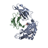
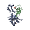
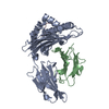
| Structure viewer | Molecule: MolmilJmol/JSmol |
|---|
- Downloads & links
-Download
| PDBx/mmCIF format | 2pcj.cif.gz | 107.8 KB | Display | PDBx/mmCIF format |
|---|---|---|---|---|
| PDB format | pdb2pcj.ent.gz | 82.1 KB | Display | PDB format |
| PDBx/mmJSON format | 2pcj.json.gz | Tree view | PDBx/mmJSON format | |
| Others |  Other downloads Other downloads |
-Validation report
| Arichive directory | https://data.pdbj.org/pub/pdb/validation_reports/pc/2pcjftp://data.pdbj.org/pub/pdb/validation_reports/pc/2pcj | HTTPS FTP |
|---|
-Related structure data
| Related structure data |  1f3oS S: Starting model for refinement |
|---|---|
| Similar structure data | |
| Other databases |


-Links
 PDBj
PDBj


- Assembly
| Deposited unit | 
| ||||||||
|---|---|---|---|---|---|---|---|---|---|
| 1 |
| ||||||||
| Unit cell |
|
-Components
| #1: Protein | Mass: 24923.045 Da / Num. of mol.: 2 Source method: isolated from a genetically manipulated source Source: (gene. exp.) Aquifex aeolicus (bacteria) / Strain: VF5 / Plasmid: pET21A / Production host: Escherichia coli (E. coli) / Strain (production host): BL-21 CONDON PLUS (DE3)-RILReferences: UniProt: O66646, Hydrolases; Acting on acid anhydrides; Acting on acid anhydrides to catalyse transmembrane movement of substances#2: Chemical | ChemComp-SO3 / | Sulfite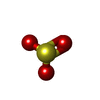  Mass: 80.063 Da / Num. of mol.: 1 / Source method: obtained synthetically / Formula: SO3 Mass: 80.063 Da / Num. of mol.: 1 / Source method: obtained synthetically / Formula: SO3#3: Water | ChemComp-HOH / | Water Mass: 18.015 Da / Num. of mol.: 452 / Source method: isolated from a natural source / Formula: H2O Mass: 18.015 Da / Num. of mol.: 452 / Source method: isolated from a natural source / Formula: H2O |
|---|
-Experimental details
-Experiment
| Experiment | Method: X-RAY DIFFRACTION / Number of used crystals: 1 |
|---|
- Sample preparation
| Crystal | Density Matthews: 2.08 Å3/Da / Density % sol: 40.87 % |
|---|---|
| Crystal grow | Temperature: 293 K / Method: vapor diffusion, sitting drop / pH: 6.4 Details: 20% isopropanol, 0.1 M Na Citrate, 16% PEG 4000, pH 6.4, VAPOR DIFFUSION, SITTING DROP, temperature 293K |
-Data collection
| Diffraction | Mean temperature: 100 K |
|---|---|
| Diffraction source | Source: SYNCHROTRON / Site: SPring-8  / Beamline: BL45PX / Wavelength: 0.9794 Å / Beamline: BL45PX / Wavelength: 0.9794 Å |
| Detector | Type: RIGAKU RAXIS V / Detector: IMAGE PLATE / Date: Dec 17, 2006 / Details: RH Coated Bent-Cyrindrical MIRROR |
| Radiation | Monochromator: SI 1 1 1 DOUBLE CRYSTAL MONOCHROMATOR / Protocol: SINGLE WAVELENGTH / Monochromatic (M) / Laue (L): M / Scattering type: x-ray |
| Radiation wavelength | Wavelength: 0.9794 Å / Relative weight: 1 |
| Reflection | Resolution: 1.7→50 Å / Num. all: 46193 / Num. obs: 46193 / % possible obs: 99.3 % / Biso Wilson estimate: 12.1 Å2 / Rmerge(I) obs: 0.041 / Rsym value: 0.044 |
| Reflection shell | Resolution: 1.7→1.76 Å / Rmerge(I) obs: 0.19 / Num. unique all: 4282 / Rsym value: 0.188 / % possible all: 93.6 |
- Processing
| Software |
| ||||||||||||||||||||
|---|---|---|---|---|---|---|---|---|---|---|---|---|---|---|---|---|---|---|---|---|---|
| Refinement | Method to determine structure: MOLECULAR REPLACEMENT Starting model: PDB ENTRY 1F3O Resolution: 1.7→45.96 Å / Rfactor Rfree error: 0.003 / Data cutoff high absF: 3052149.04 / Data cutoff low absF: 0 / Isotropic thermal model: RESTRAINED / Cross valid method: THROUGHOUT / σ(F): 0 / Stereochemistry target values: Engh & Huber
| ||||||||||||||||||||
| Solvent computation | Solvent model: FLAT MODEL / Bsol: 54.9807 Å2 / ksol: 0.369103 e/Å3 | ||||||||||||||||||||
| Displacement parameters | Biso mean: 17.6 Å2
| ||||||||||||||||||||
| Refine analyze |
| ||||||||||||||||||||
| Refinement step | Cycle: LAST / Resolution: 1.7→45.96 Å
| ||||||||||||||||||||
| Refine LS restraints |
| ||||||||||||||||||||
| LS refinement shell | Resolution: 1.7→1.78 Å / Rfactor Rfree error: 0.011 / Total num. of bins used: 8
| ||||||||||||||||||||
| Xplor file |
|
