 Movie
Movie Controller


+ Open data

- Basic information
| Entry | Database: PDB / ID: 1f3o | ||||||
|---|---|---|---|---|---|---|---|
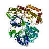
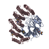
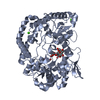
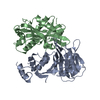
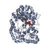
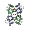
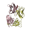
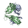
| Title | Crystal structure of MJ0796 ATP-binding cassette | ||||||
 Components Components | HYPOTHETICAL ABC TRANSPORTER ATP-BINDING PROTEIN MJ0796 | ||||||
 Keywords Keywords |  STRUCTURAL GENOMICS / Transporter STRUCTURAL GENOMICS / Transporter | ||||||
| Function / homology |  Function and homology information Function and homology informationtransmembrane transporter activity / transmembrane transport / ATP hydrolysis activity / ATP binding / plasma membraneSimilarity search - Function | ||||||
| Biological species |   Methanocaldococcus jannaschii (archaea) Methanocaldococcus jannaschii (archaea) | ||||||
| Method | X-RAY DIFFRACTION / SYNCHROTRON / Resolution: 2.7 Å | ||||||
 Authors Authors | Yuan, Y.-R. / Hunt, J.F. | ||||||
 Citation Citation | Journal: J.Biol.Chem. / Year: 2001 Title: The crystal structure of the MJ0796 ATP-binding cassette. Implications for the structural consequences of ATP hydrolysis in the active site of an ABC transporter. Authors: Yuan, Y.R. / Blecker, S. / Martsinkevich, O. / Millen, L. / Thomas, P.J. / Hunt, J.F. | ||||||
| History |
|
- Structure visualization
| Structure viewer | Molecule: MolmilJmol/JSmol |
|---|
- Downloads & links
-Download
| PDBx/mmCIF format | 1f3o.cif.gz | 56.9 KB | Display | PDBx/mmCIF format |
|---|---|---|---|---|
| PDB format | pdb1f3o.ent.gz | 45.7 KB | Display | PDB format |
| PDBx/mmJSON format | 1f3o.json.gz | Tree view | PDBx/mmJSON format | |
| Others |  Other downloads Other downloads |
-Validation report
| Arichive directory | https://data.pdbj.org/pub/pdb/validation_reports/f3/1f3oftp://data.pdbj.org/pub/pdb/validation_reports/f3/1f3o | HTTPS FTP |
|---|
-Related structure data
| Similar structure data |
|---|
-Links
 PDBj
PDBj

- Assembly
| Deposited unit | 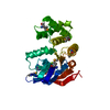
| ||||||||
|---|---|---|---|---|---|---|---|---|---|
| 1 | 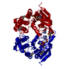
| ||||||||
| Unit cell |
| ||||||||
| Details | The biological assembly is a dimer |
-Components
| #1: Protein | Mass: 26931.941 Da / Num. of mol.: 1 Source method: isolated from a genetically manipulated source Source: (gene. exp.) Methanocaldococcus jannaschii (archaea)Plasmid: PET28A / Gene (production host): GENOMIC DNA / Production host:  Escherichia coli (E. coli) / References: UniProt: Q58206 Escherichia coli (E. coli) / References: UniProt: Q58206 |
|---|---|
| #2: Chemical | ChemComp-MG /   Mass: 24.305 Da / Num. of mol.: 1 / Source method: obtained synthetically / Formula: Mg Mass: 24.305 Da / Num. of mol.: 1 / Source method: obtained synthetically / Formula: Mg |
| #3: Chemical | ChemComp-ADP / Adenosine diphosphate  Mass: 427.201 Da / Num. of mol.: 1 / Source method: obtained synthetically / Formula: C10H15N5O10P2 / Comment: ADP, energy-carrying molecule*YM Mass: 427.201 Da / Num. of mol.: 1 / Source method: obtained synthetically / Formula: C10H15N5O10P2 / Comment: ADP, energy-carrying molecule*YM |
| #4: Water | ChemComp-HOH / Water Mass: 18.015 Da / Num. of mol.: 64 / Source method: isolated from a natural source / Formula: H2O Mass: 18.015 Da / Num. of mol.: 64 / Source method: isolated from a natural source / Formula: H2O |
-Experimental details
-Experiment
| Experiment | Method: X-RAY DIFFRACTION / Number of used crystals: 1 |
|---|
- Sample preparation
| Crystal | Density Matthews: 2.12 Å3/Da / Density % sol: 41.98 % | ||||||||||||||||||||||||||||||||||||||||||||||||
|---|---|---|---|---|---|---|---|---|---|---|---|---|---|---|---|---|---|---|---|---|---|---|---|---|---|---|---|---|---|---|---|---|---|---|---|---|---|---|---|---|---|---|---|---|---|---|---|---|---|
| Crystal grow | Temperature: 294 K / Method: vapor diffusion, hanging drop / pH: 6.8 Details: PEG 4000, Ammonium Acetate, Glycerol, ADP, Magnesium Chloride, pH 6.8, VAPOR DIFFUSION, HANGING DROP, temperature 21K | ||||||||||||||||||||||||||||||||||||||||||||||||
| Crystal grow | *PLUS pH: 7.6 | ||||||||||||||||||||||||||||||||||||||||||||||||
| Components of the solutions | *PLUS
|
-Data collection
| Diffraction | Mean temperature: 130 K |
|---|---|
| Diffraction source | Source: SYNCHROTRON / Site: NSLS  / Beamline: X12C / Wavelength: 0.932 / Beamline: X12C / Wavelength: 0.932 |
| Detector | Type: BRANDEIS / Detector: CCD / Date: Apr 20, 2000 |
| Radiation | Protocol: SINGLE WAVELENGTH / Monochromatic (M) / Laue (L): M / Scattering type: x-ray |
| Radiation wavelength | Wavelength: 0.932 Å / Relative weight: 1 |
| Reflection | Resolution: 2.7→30 Å / Num. all: 25396 / Num. obs: 24520 / % possible obs: 93.5 % / Observed criterion σ(F): 1 / Observed criterion σ(I): 3 / Redundancy: 4.42 % / Biso Wilson estimate: 24.1 Å2 / Rmerge(I) obs: 0.146 / Net I/σ(I): 5.5 |
| Reflection shell | Resolution: 2.7→2.82 Å / Redundancy: 3.9 % / % possible all: 74.5 |
- Processing
| Software |
| ||||||||||||||||||||||||||||||||||||||||
|---|---|---|---|---|---|---|---|---|---|---|---|---|---|---|---|---|---|---|---|---|---|---|---|---|---|---|---|---|---|---|---|---|---|---|---|---|---|---|---|---|---|
| Refinement | Resolution: 2.7→30 Å / Rfactor Rfree error: 0.011 / Data cutoff high absF: 1000000 / Data cutoff low absF: 0.01 / Isotropic thermal model: RESTRAINED / Cross valid method: THROUGHOUT / σ(F): 1 / σ(I): 1 / Stereochemistry target values: X-Plor 3.851
| ||||||||||||||||||||||||||||||||||||||||
| Displacement parameters | Biso mean: 41.4 Å2
| ||||||||||||||||||||||||||||||||||||||||
| Refine analyze |
| ||||||||||||||||||||||||||||||||||||||||
| Refinement step | Cycle: LAST / Resolution: 2.7→30 Å
| ||||||||||||||||||||||||||||||||||||||||
| Refine LS restraints |
| ||||||||||||||||||||||||||||||||||||||||
| LS refinement shell | Resolution: 2.7→2.82 Å / Rfactor Rfree error: 0.057 / Total num. of bins used: 8
| ||||||||||||||||||||||||||||||||||||||||
| Software | *PLUS Name: X-PLOR(ONLINE) / Version: 3.851 / Classification: refinement | ||||||||||||||||||||||||||||||||||||||||
| Refinement | *PLUS σ(F): 1 / % reflection Rfree: 11.1 % / Rfactor obs: 0.204 | ||||||||||||||||||||||||||||||||||||||||
| Solvent computation | *PLUS | ||||||||||||||||||||||||||||||||||||||||
| Displacement parameters | *PLUS Biso mean: 41.4 Å2 | ||||||||||||||||||||||||||||||||||||||||
| Refine LS restraints | *PLUS
| ||||||||||||||||||||||||||||||||||||||||
| LS refinement shell | *PLUS Rfactor Rfree: 0.456 / % reflection Rfree: 10.7 % / Rfactor Rwork: 0.368 |
