 Movie
Movie Controller

[English] 日本語
 Yorodumi
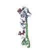
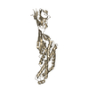
- PDB-2p28: Structure of the PHE2 and PHE3 fragments of the integrin beta2 subunit -
+ Open data

- Basic information
| Entry | Database: PDB / ID: 2p28 | ||||||
|---|---|---|---|---|---|---|---|
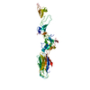
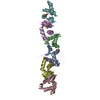
| Title | Structure of the PHE2 and PHE3 fragments of the integrin beta2 subunit | ||||||
 Components Components | (Integrin beta-2) x 2 | ||||||
 Keywords Keywords |  CELL ADHESION / integrin beta2 subunit / hybrid domain / PSI domain / I-EGF domains CELL ADHESION / integrin beta2 subunit / hybrid domain / PSI domain / I-EGF domains | ||||||
| Function / homology |  Function and homology information Function and homology informationintegrin alphaX-beta2 complex / integrin alphaM-beta2 complex / positive regulation of prostaglandin-E synthase activity / positive regulation of neutrophil degranulation / integrin alphaL-beta2 complex / ICAM-3 receptor activity / complement component C3b binding / neutrophil migration / Toll Like Receptor 4 (TLR4) Cascade / negative regulation of dopamine metabolic process ...integrin alphaX-beta2 complex / integrin alphaM-beta2 complex / positive regulation of prostaglandin-E synthase activity / positive regulation of neutrophil degranulation / integrin alphaL-beta2 complex / ICAM-3 receptor activity / complement component C3b binding / neutrophil migration / Toll Like Receptor 4 (TLR4) Cascade / negative regulation of dopamine metabolic process / cell-cell adhesion via plasma-membrane adhesion molecules / heterotypic cell-cell adhesion / integrin complex / positive regulation of leukocyte adhesion to vascular endothelial cell / cell adhesion mediated by integrin / leukocyte cell-cell adhesion / phagocytosis, engulfment / receptor clustering / amyloid-beta clearance / endodermal cell differentiation / plasma membrane raft / tertiary granule membrane / ficolin-1-rich granule membrane / cellular response to low-density lipoprotein particle stimulus / positive regulation of protein targeting to membrane / Integrin cell surface interactions / specific granule membrane / regulation of peptidyl-tyrosine phosphorylation / heat shock protein binding / cell adhesion molecule binding / receptor-mediated endocytosis / neutrophil chemotaxis / cell-matrix adhesion / positive regulation of superoxide anion generation / integrin-mediated signaling pathway / Cell surface interactions at the vascular wall / microglial cell activation / receptor internalization / cell-cell adhesion / Immunoregulatory interactions between a Lymphoid and a non-Lymphoid cell / extracellular vesicle / integrin binding / cell-cell signaling / amyloid-beta binding / regulation of cell shape / Interleukin-4 and Interleukin-13 signaling / receptor complex / cell adhesion / inflammatory response / external side of plasma membrane / focal adhesion / apoptotic process / Neutrophil degranulation / protein kinase binding / cell surface / extracellular exosome / membrane / metal ion binding / plasma membraneSimilarity search - Function | ||||||
| Biological species |  Homo sapiens (human) Homo sapiens (human) | ||||||
| Method | X-RAY DIFFRACTION / SYNCHROTRON / MOLECULAR REPLACEMENT / Resolution: 2.2 Å | ||||||
 Authors Authors | Shi, M. / Foo, S.Y. / Tan, S.M. / Mitchell, E.P. / Law, S.K.A. / Lescar, J. | ||||||
 Citation Citation | Journal: J.Biol.Chem. / Year: 2007 Title: A structural hypothesis for the transition between bent and extended conformations of the leukocyte beta2 integrins Authors: Shi, M. / Foo, S.Y. / Tan, S.M. / Mitchell, E.P. / Law, S.K.A. / Lescar, J. | ||||||
| History |
|
- Structure visualization
| Structure viewer | Molecule: MolmilJmol/JSmol |
|---|
- Downloads & links
-Download
| PDBx/mmCIF format | 2p28.cif.gz | 79.8 KB | Display | PDBx/mmCIF format |
|---|---|---|---|---|
| PDB format | pdb2p28.ent.gz | 58.7 KB | Display | PDB format |
| PDBx/mmJSON format | 2p28.json.gz | Tree view | PDBx/mmJSON format | |
| Others |  Other downloads Other downloads |
-Validation report
| Arichive directory | https://data.pdbj.org/pub/pdb/validation_reports/p2/2p28ftp://data.pdbj.org/pub/pdb/validation_reports/p2/2p28 | HTTPS FTP |
|---|
-Related structure data
| Related structure data | 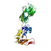 2p26C  1l3yS 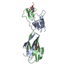 1yukS C: citing same article ( S: Starting model for refinement |
|---|---|
| Similar structure data |
-Links
 PDBj
PDBj


- Assembly
| Deposited unit | 
| ||||||||
|---|---|---|---|---|---|---|---|---|---|
| 1 |
| ||||||||
| Unit cell |
|
-Components
| #1: Protein | Mass: 11037.438 Da / Num. of mol.: 1 / Fragment: PHE2 fragment Source method: isolated from a genetically manipulated source Source: (gene. exp.) Homo sapiens (human) / Plasmid: pIRES2-EGFP / Cell line (production host): HEK293_Gnti / Production host: Homo sapiens (human) / References: UniProt: P05107 |
|---|---|
| #2: Protein | Mass: 23879.072 Da / Num. of mol.: 1 / Fragment: PHE3 fragment Source method: isolated from a genetically manipulated source Source: (gene. exp.) Homo sapiens (human) / Plasmid: pIRES2-EGFP / Cell line (production host): HEK293_Gnti / Production host: Homo sapiens (human) / References: UniProt: P05107 |
| #3: Sugar | ChemComp-NAG / N-Acetylglucosamine  Type: D-saccharide, beta linking / Mass: 221.208 Da / Num. of mol.: 1 Type: D-saccharide, beta linking / Mass: 221.208 Da / Num. of mol.: 1Source method: isolated from a genetically manipulated source Formula: C8H15NO6 |
| #4: Water | ChemComp-HOH / Water Mass: 18.015 Da / Num. of mol.: 199 / Source method: isolated from a natural source / Formula: H2O Mass: 18.015 Da / Num. of mol.: 199 / Source method: isolated from a natural source / Formula: H2O |
-Experimental details
-Experiment
| Experiment | Method: X-RAY DIFFRACTION / Number of used crystals: 1 |
|---|
- Sample preparation
| Crystal | Density Matthews: 2.39 Å3/Da / Density % sol: 48.58 % |
|---|---|
| Crystal grow | Temperature: 291 K / Method: vapor diffusion, hanging drop Details: 0.2M ammonium sulphate, 15% PEG4000, 5% isopropanol, VAPOR DIFFUSION, HANGING DROP, temperature 291K |
-Data collection
| Diffraction | Mean temperature: 100 K |
|---|---|
| Diffraction source | Source: SYNCHROTRON / Site: ESRF  / Beamline: ID23-1 / Wavelength: 0.976 Å / Beamline: ID23-1 / Wavelength: 0.976 Å |
| Detector | Type: ADSC QUANTUM 4 / Detector: CCD / Date: Sep 25, 2006 |
| Radiation | Protocol: SINGLE WAVELENGTH / Monochromatic (M) / Laue (L): M / Scattering type: x-ray |
| Radiation wavelength | Wavelength: 0.976 Å / Relative weight: 1 |
| Reflection | Resolution: 2.2→25 Å / Num. obs: 18866 |
| Reflection shell | Resolution: 2.2→2.257 Å |
- Processing
| Software |
| ||||||||||||||||||||||||||||||||||||||||||||||||||||||||||||||||||||||||||||||||||||||||||||||||||||
|---|---|---|---|---|---|---|---|---|---|---|---|---|---|---|---|---|---|---|---|---|---|---|---|---|---|---|---|---|---|---|---|---|---|---|---|---|---|---|---|---|---|---|---|---|---|---|---|---|---|---|---|---|---|---|---|---|---|---|---|---|---|---|---|---|---|---|---|---|---|---|---|---|---|---|---|---|---|---|---|---|---|---|---|---|---|---|---|---|---|---|---|---|---|---|---|---|---|---|---|---|---|
| Refinement | Method to determine structure: MOLECULAR REPLACEMENT Starting model: 1YUK, 1L3Y Resolution: 2.2→25 Å / Cor.coef. Fo:Fc: 0.912 / Cor.coef. Fo:Fc free: 0.9 / SU B: 7.734 / SU ML: 0.196 / TLS residual ADP flag: UNVERIFIED / Cross valid method: THROUGHOUT / σ(F): 0 / ESU R: 0.341 / ESU R Free: 0.262 / Stereochemistry target values: MAXIMUM LIKELIHOOD / Details: HYDROGENS HAVE BEEN ADDED IN THE RIDING POSITIONS
| ||||||||||||||||||||||||||||||||||||||||||||||||||||||||||||||||||||||||||||||||||||||||||||||||||||
| Solvent computation | Ion probe radii: 0.8 Å / Shrinkage radii: 0.8 Å / VDW probe radii: 1.4 Å / Solvent model: BABINET MODEL WITH MASK | ||||||||||||||||||||||||||||||||||||||||||||||||||||||||||||||||||||||||||||||||||||||||||||||||||||
| Displacement parameters | Biso mean: 22.65 Å2
| ||||||||||||||||||||||||||||||||||||||||||||||||||||||||||||||||||||||||||||||||||||||||||||||||||||
| Refinement step | Cycle: LAST / Resolution: 2.2→25 Å
| ||||||||||||||||||||||||||||||||||||||||||||||||||||||||||||||||||||||||||||||||||||||||||||||||||||
| Refine LS restraints |
| ||||||||||||||||||||||||||||||||||||||||||||||||||||||||||||||||||||||||||||||||||||||||||||||||||||
| LS refinement shell | Resolution: 2.2→2.257 Å / Total num. of bins used: 20
| ||||||||||||||||||||||||||||||||||||||||||||||||||||||||||||||||||||||||||||||||||||||||||||||||||||
| Refinement TLS params. | Method: refined / Refine-ID: X-RAY DIFFRACTION
| ||||||||||||||||||||||||||||||||||||||||||||||||||||||||||||||||||||||||||||||||||||||||||||||||||||
| Refinement TLS group | Refine-ID: X-RAY DIFFRACTION / Selection: ALL
|
