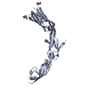
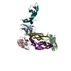
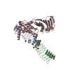
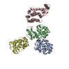
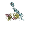
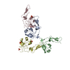
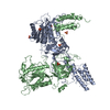
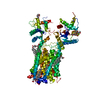
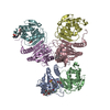
[English] 日本語
Yorodumi
- PDB-2oz4: Structural Plasticity in IgSF Domain 4 of ICAM-1 Mediates Cell Su... -

+
Open data


ID or keywords:

Loading...

-
Basic information

Entry
Database: PDB / ID: 2oz4
TitleStructural Plasticity in IgSF Domain 4 of ICAM-1 Mediates Cell Surface Dimerization
Components
  • FAB FRAGMENT LIGHT CHAINFragment antigen-binding
  • FAB FRAGMENT, HEAVY CHAINFragment antigen-binding
  • Intercellular adhesion molecule 1
KeywordsCELL ADHESION / IGSF DOMAIN / STRUCTURAL PLASTICITY / CELL-SURFACE DIMERIZATION
Function / homology
Function and homology information


response to sulfur dioxide / response to gonadotropin / : / regulation of leukocyte mediated cytotoxicity / T cell extravasation / positive regulation of cellular extravasation / regulation of ruffle assembly / establishment of Sertoli cell barrier / T cell antigen processing and presentation / T cell activation via T cell receptor contact with antigen bound to MHC molecule on antigen presenting cell ...response to sulfur dioxide / response to gonadotropin / : / regulation of leukocyte mediated cytotoxicity / T cell extravasation / positive regulation of cellular extravasation / regulation of ruffle assembly / establishment of Sertoli cell barrier / T cell antigen processing and presentation / T cell activation via T cell receptor contact with antigen bound to MHC molecule on antigen presenting cell / membrane to membrane docking / acute inflammatory response to antigenic stimulus / establishment of endothelial intestinal barrier / response to xenobiotic stimulus => GO:0009410 / adhesion of symbiont to host / positive regulation of B cell activation / phagocytosis, recognition / cellular response to interleukin-6 / establishment of endothelial barrier / response to copper ion / positive regulation of leukocyte adhesion to vascular endothelial cell / cell adhesion mediated by integrin / heterophilic cell-cell adhesion via plasma membrane cell adhesion molecules / phagocytosis, engulfment / leukocyte cell-cell adhesion / response to ionizing radiation / leukocyte migration / positive regulation of actin filament polymerization / immunoglobulin complex, circulating / cellular response to alkaloid / immunoglobulin receptor binding / plasma membrane => GO:0005886 / Interleukin-10 signaling / cellular response to nutrient levels / negative regulation of calcium ion transport / immunological synapse / regulation of immune response / response to amino acid / Integrin cell surface interactions / cellular response to interleukin-1 / negative regulation of endothelial cell apoptotic process / type II interferon-mediated signaling pathway / complement activation, classical pathway / negative regulation of extrinsic apoptotic signaling pathway via death domain receptors / ovarian follicle development / positive regulation of vasoconstriction / response to amphetamine / antigen binding / cellular response to leukemia inhibitory factor / extracellular matrix organization / cellular response to dexamethasone stimulus / cellular response to glucose stimulus / sensory perception of sound / B cell receptor signaling pathway / response to insulin / positive regulation of GTPase activity / cytokine-mediated signaling pathway / cellular response to amyloid-beta / positive regulation of nitric oxide biosynthetic process / Immunoregulatory interactions between a Lymphoid and a non-Lymphoid cell / Interferon gamma signaling / positive regulation of peptidyl-tyrosine phosphorylation / transmembrane signaling receptor activity / integrin binding / virus receptor activity / signaling receptor activity / cellular response to tumor necrosis factor / positive regulation of NF-kappaB transcription factor activity / regulation of cell shape / cellular response to hypoxia / collagen-containing extracellular matrix / response to ethanol / Interleukin-4 and Interleukin-13 signaling / cellular response to lipopolysaccharide / receptor-mediated virion attachment to host cell / positive regulation of ERK1 and ERK2 cascade / cell adhesion / defense response to bacterium / membrane raft / external side of plasma membrane / focal adhesion / innate immune response / cell surface / extracellular space / extracellular exosome / membrane / plasma membrane
Similarity search - Function
: / ICAM-1/3/5, D2 domain / Intercellular adhesion molecule / Intercellular adhesion molecule, N-terminal / : / Intercellular adhesion molecule (ICAM), N-terminal domain / Intercellular adhesion molecule/vascular cell adhesion molecule, N-terminal / Immunoglobulin domain / Immunoglobulin V-Type / Immunoglobulin V-set domain ...: / ICAM-1/3/5, D2 domain / Intercellular adhesion molecule / Intercellular adhesion molecule, N-terminal / : / Intercellular adhesion molecule (ICAM), N-terminal domain / Intercellular adhesion molecule/vascular cell adhesion molecule, N-terminal / Immunoglobulin domain / Immunoglobulin V-Type / Immunoglobulin V-set domain / Immunoglobulin V-set domain / Immunoglobulin subtype / Immunoglobulin / Ig-like domain profile. / Immunoglobulin-like domain / Immunoglobulin-like domain superfamily / Immunoglobulins / Immunoglobulin-like fold / Immunoglobulin-like / Sandwich / Mainly Beta
Similarity search - Domain/homology
Ig heavy chain V region J558 / Intercellular adhesion molecule 1 / : / Intercellular adhesion molecule 1
Similarity search - Component
Biological speciesHomo sapiens (human)
Mus musculus (house mouse)
MethodX-RAY DIFFRACTION / SYNCHROTRON / MOLECULAR REPLACEMENT / Resolution: 2.7 Å
AuthorsChen, X. / Kim, T.D. / Carman, C.V. / Mi, L. / Song, G. / Springer, T.A.
CitationJournal: Proc.Natl.Acad.Sci.Usa / Year: 2007
Title: Structural plasticity in Ig superfamily domain 4 of ICAM-1 mediates cell surface dimerization.
Authors: Chen, X. / Kim, T.D. / Carman, C.V. / Mi, L.Z. / Song, G. / Springer, T.A.
History
DepositionFeb 23, 2007Deposition site: RCSB / Processing site: RCSB
Revision 1.0Oct 16, 2007Provider: repository / Type: Initial release
Revision 1.1Jul 13, 2011Group: Advisory / Version format compliance
Revision 2.0Jul 29, 2020Group: Atomic model / Data collection ...Atomic model / Data collection / Derived calculations / Structure summary
Category: atom_site / chem_comp ...atom_site / chem_comp / entity / pdbx_branch_scheme / pdbx_chem_comp_identifier / pdbx_entity_branch / pdbx_entity_branch_descriptor / pdbx_entity_branch_link / pdbx_entity_branch_list / pdbx_entity_nonpoly / pdbx_nonpoly_scheme / pdbx_struct_assembly_gen / pdbx_struct_conn_angle / pdbx_struct_special_symmetry / struct_asym / struct_conn / struct_site / struct_site_gen
Item: _atom_site.auth_asym_id / _atom_site.auth_seq_id ..._atom_site.auth_asym_id / _atom_site.auth_seq_id / _atom_site.label_asym_id / _atom_site.label_entity_id / _chem_comp.name / _chem_comp.type / _entity.formula_weight / _entity.pdbx_description / _entity.pdbx_number_of_molecules / _entity.src_method / _entity.type / _pdbx_struct_assembly_gen.asym_id_list / _pdbx_struct_conn_angle.ptnr1_auth_seq_id / _pdbx_struct_conn_angle.ptnr1_label_asym_id / _pdbx_struct_conn_angle.ptnr2_label_asym_id / _pdbx_struct_conn_angle.ptnr3_auth_seq_id / _pdbx_struct_conn_angle.ptnr3_label_asym_id / _pdbx_struct_conn_angle.value / _pdbx_struct_special_symmetry.label_asym_id / _struct_conn.pdbx_dist_value / _struct_conn.pdbx_leaving_atom_flag / _struct_conn.pdbx_role / _struct_conn.ptnr1_auth_asym_id / _struct_conn.ptnr1_auth_seq_id / _struct_conn.ptnr1_label_asym_id / _struct_conn.ptnr1_label_seq_id / _struct_conn.ptnr2_auth_asym_id / _struct_conn.ptnr2_auth_seq_id / _struct_conn.ptnr2_label_asym_id
Description: Carbohydrate remediation / Provider: repository / Type: Remediation
Revision 2.1Aug 30, 2023Group: Data collection / Database references ...Data collection / Database references / Refinement description / Structure summary
Category: chem_comp / chem_comp_atom ...chem_comp / chem_comp_atom / chem_comp_bond / database_2 / pdbx_initial_refinement_model
Item: _chem_comp.pdbx_synonyms / _database_2.pdbx_DOI / _database_2.pdbx_database_accession

-
Structure visualization

Structure viewerMolecule:
MolmilJmol/JSmol

Downloads & links

-
Assembly

Deposited unit
A: Intercellular adhesion molecule 1
L: FAB FRAGMENT LIGHT CHAIN
H: FAB FRAGMENT, HEAVY CHAIN
hetero molecules


Theoretical massNumber of molelcules
Total (without water)76,42612
Polymers75,0493
Non-polymers1,3779
Water4,972276
1


  • Idetical with deposited unit
  • defined by author&software
TypeNameSymmetry operationNumber
identity operation1_555x,y,z1
MethodPISA
Unit cell
Length a, b, c (Å)185.396, 69.342, 88.173
Angle α, β, γ (deg.)90.00, 112.76, 90.00
Int Tables number5
Space group name H-MC121
Components on special symmetry positions
IDModelComponents
11L-2002-

ZN

21L-3081-

HOH

-
Components

-
Protein , 1 types, 1 molecules A

#1: Protein Intercellular adhesion molecule 1 /


Mass: 28682.322 Da / Num. of mol.: 1
Source method: isolated from a genetically manipulated source
Source: (gene. exp.) Homo sapiens (human) / Gene: ICAM1 / Production host: Cricetulus griseus (Chinese hamster) / Strain (production host): CHINESE HAMSTER CHO.LEC3.2.8.1 / References: UniProt: Q5NKV7, UniProt: P05362*PLUS

-
Antibody , 2 types, 2 molecules LH

#2: Antibody FAB FRAGMENT LIGHT CHAIN / Fragment antigen-binding


Mass: 23592.920 Da / Num. of mol.: 1 / Source method: isolated from a natural source / Source: (natural) Mus musculus (house mouse) / References: UniProt: Q58EU4
#3: Antibody FAB FRAGMENT, HEAVY CHAIN / Fragment antigen-binding


Mass: 22773.498 Da / Num. of mol.: 1 / Source method: isolated from a natural source / Source: (natural) Mus musculus (house mouse) / References: UniProt: P01757

-
Sugars , 2 types, 3 molecules

#4: Polysaccharide alpha-L-fucopyranose-(1-6)-2-acetamido-2-deoxy-beta-D-glucopyranose


Type: oligosaccharide / Mass: 367.349 Da / Num. of mol.: 1
Source method: isolated from a genetically manipulated source
DescriptorTypeProgram
LFucpa1-6DGlcpNAcb1-Glycam Condensed SequenceGMML 1.0
WURCS=2.0/2,2,1/[a2122h-1b_1-5_2*NCC/3=O][a1221m-1a_1-5]/1-2/a6-b1WURCSPDB2Glycan 1.1.0
[]{[(4+1)][b-D-GlcpNAc]{[(6+1)][a-L-Fucp]{}}}LINUCSPDB-CARE
#5: Sugar ChemComp-NAG / 2-acetamido-2-deoxy-beta-D-glucopyranose / N-acetyl-beta-D-glucosamine / 2-acetamido-2-deoxy-beta-D-glucose / 2-acetamido-2-deoxy-D-glucose / 2-acetamido-2-deoxy-glucose / N-ACETYL-D-GLUCOSAMINE / N-Acetylglucosamine


Type: D-saccharide, beta linking / Mass: 221.208 Da / Num. of mol.: 2 / Source method: obtained synthetically / Formula: C8H15NO6
IdentifierTypeProgram
DGlcpNAcbCONDENSED IUPAC CARBOHYDRATE SYMBOLGMML 1.0
N-acetyl-b-D-glucopyranosamineCOMMON NAMEGMML 1.0
b-D-GlcpNAcIUPAC CARBOHYDRATE SYMBOLPDB-CARE 1.0
GlcNAcSNFG CARBOHYDRATE SYMBOLGMML 1.0

-
Non-polymers , 4 types, 282 molecules

#6: Chemical ChemComp-SO4 / SULFATE ION / Sulfate


Mass: 96.063 Da / Num. of mol.: 2 / Source method: obtained synthetically / Formula: SO4
#7: Chemical ChemComp-ZN / ZINC ION


Mass: 65.409 Da / Num. of mol.: 2 / Source method: obtained synthetically / Formula: Zn
#8: Chemical ChemComp-TRS / 2-AMINO-2-HYDROXYMETHYL-PROPANE-1,3-DIOL / TRIS BUFFER / Tris


Mass: 122.143 Da / Num. of mol.: 2 / Source method: obtained synthetically / Formula: C4H12NO3 / Comment: pH buffer*YM
#9: Water ChemComp-HOH / water / Water


Mass: 18.015 Da / Num. of mol.: 276 / Source method: isolated from a natural source / Formula: H2O

-
Experimental details

-
Experiment

ExperimentMethod: X-RAY DIFFRACTION / Number of used crystals: 1

-
Sample preparation

CrystalDensity Matthews: 3.49 Å3/Da / Density % sol: 64.78 %
Crystal growTemperature: 293 K / Method: vapor diffusion, hanging drop / pH: 7.5
Details: 1.5M LITHIUM SULFATE, 0.1M HEPES, pH 7.5, VAPOR DIFFUSION, HANGING DROP, temperature 293K

-
Data collection

DiffractionMean temperature: 100 K
Diffraction sourceSource: SYNCHROTRON / Site: APS / Beamline: 19-ID / Wavelength: 0.97949 Å
DetectorType: ADSC QUANTUM 315 / Detector: CCD / Date: Jan 1, 2006
RadiationMonochromator: Rosenbaum-Rock high-resolution double-crystal monochromator
Protocol: SINGLE WAVELENGTH / Monochromatic (M) / Laue (L): M / Scattering type: x-ray
Radiation wavelengthWavelength: 0.97949 Å / Relative weight: 1
ReflectionResolution: 2.7→30 Å / Num. all: 26015 / Num. obs: 26015 / % possible obs: 90.6 % / Observed criterion σ(I): 0 / Redundancy: 3.5 % / Rmerge(I) obs: 0.087 / Rsym value: 0.087 / Net I/σ(I): 11.1
Reflection shellResolution: 2.7→2.8 Å / Redundancy: 2.7 % / Rmerge(I) obs: 0.543 / Mean I/σ(I) obs: 1.2 / Rsym value: 0.543 / % possible all: 58.1

-
Processing

Software
NameVersionClassification
REFMAC5.2.0019refinement
HKL-2000data collection
HKL-2000data reduction
HKL-2000data scaling
MOLREPphasing
RefinementMethod to determine structure: MOLECULAR REPLACEMENT
Starting model: pdb entries 1F11, 1P53
Resolution: 2.7→28.5 Å / Cor.coef. Fo:Fc: 0.95 / Cor.coef. Fo:Fc free: 0.923 / SU B: 26.501 / SU ML: 0.288 / TLS residual ADP flag: LIKELY RESIDUAL / Isotropic thermal model: overall / Cross valid method: THROUGHOUT / ESU R: 1.353 / ESU R Free: 0.346 / Stereochemistry target values: MAXIMUM LIKELIHOOD / Details: HYDROGENS HAVE BEEN ADDED IN THE RIDING POSITIONS
RfactorNum. reflection% reflectionSelection details
Rfree0.253 2559 9.9 %RANDOM
Rwork0.204 ---
obs0.209 23400 100 %-
all-25959 --
Solvent computationIon probe radii: 0.8 Å / Shrinkage radii: 0.8 Å / VDW probe radii: 1.4 Å / Solvent model: MASK
Displacement parametersBiso mean: 63.34 Å2
Baniso -1Baniso -2Baniso -3
1--5.52 Å20 Å2-4.12 Å2
2--6.95 Å20 Å2
3----4.62 Å2
Refine analyzeLuzzati coordinate error obs: 0.455 Å
Refinement stepCycle: LAST / Resolution: 2.7→28.5 Å
ProteinNucleic acidLigandSolventTotal
Num. atoms5234 0 80 276 5590
Refine LS restraints
Refine-IDTypeDev idealDev ideal targetNumber
X-RAY DIFFRACTIONr_bond_refined_d0.0050.0225449
X-RAY DIFFRACTIONr_bond_other_d0.0010.023605
X-RAY DIFFRACTIONr_angle_refined_deg0.9031.9747446
X-RAY DIFFRACTIONr_angle_other_deg0.65638848
X-RAY DIFFRACTIONr_dihedral_angle_1_deg4.965685
X-RAY DIFFRACTIONr_dihedral_angle_2_deg30.83124.72214
X-RAY DIFFRACTIONr_dihedral_angle_3_deg10.59915863
X-RAY DIFFRACTIONr_dihedral_angle_4_deg12.7881525
X-RAY DIFFRACTIONr_chiral_restr0.0910.2867
X-RAY DIFFRACTIONr_gen_planes_refined0.0020.026001
X-RAY DIFFRACTIONr_gen_planes_other0.0010.021008
X-RAY DIFFRACTIONr_nbd_refined0.1490.2864
X-RAY DIFFRACTIONr_nbd_other0.1640.23592
X-RAY DIFFRACTIONr_nbtor_refined0.1630.22617
X-RAY DIFFRACTIONr_nbtor_other0.0770.22975
X-RAY DIFFRACTIONr_xyhbond_nbd_refined0.0910.2272
X-RAY DIFFRACTIONr_xyhbond_nbd_other
X-RAY DIFFRACTIONr_metal_ion_refined0.090.21
X-RAY DIFFRACTIONr_metal_ion_other
X-RAY DIFFRACTIONr_symmetry_vdw_refined0.0930.216
X-RAY DIFFRACTIONr_symmetry_vdw_other0.1530.245
X-RAY DIFFRACTIONr_symmetry_hbond_refined0.0880.215
X-RAY DIFFRACTIONr_symmetry_hbond_other
X-RAY DIFFRACTIONr_symmetry_metal_ion_refined0.0660.22
X-RAY DIFFRACTIONr_symmetry_metal_ion_other
X-RAY DIFFRACTIONr_mcbond_it0.62734460
X-RAY DIFFRACTIONr_mcbond_other0.01631373
X-RAY DIFFRACTIONr_mcangle_it0.897185624
X-RAY DIFFRACTIONr_scbond_it0.4932333
X-RAY DIFFRACTIONr_scangle_it0.865181822
X-RAY DIFFRACTIONr_rigid_bond_restr
X-RAY DIFFRACTIONr_sphericity_free
X-RAY DIFFRACTIONr_sphericity_bonded
LS refinement shellResolution: 2.7→2.77 Å / Total num. of bins used: 20
RfactorNum. reflection% reflection
Rfree0.484 103 -
Rwork0.453 1090 -
obs--100 %
Refinement TLS params.

Method: refined / Refine-ID: X-RAY DIFFRACTION

IDL112)L122)L132)L222)L232)L332)S11 (Å °)S12 (Å °)S13 (Å °)S21 (Å °)S22 (Å °)S23 (Å °)S31 (Å °)S32 (Å °)S33 (Å °)T112)T122)T132)T222)T232)T332)Origin x (Å)Origin y (Å)Origin z (Å)
13.7888-2.20242.58483.2977-1.63365.4443-0.1355-0.02560.6613-0.08650.0192-0.0697-0.70920.23890.1163-0.307-0.13570.0182-0.1784-0.0611-0.1192-64.156110.117262.9419
29.48232.56161.7373.7392-0.13135.39140.0955-0.26520.0824-0.0955-0.13830.30020.47060.15810.0429-0.2670.00050.0304-0.2413-0.0516-0.3925-64.1176-12.626265.1816
38.46337.67951.738613.12940.26893.81390.19650.44450.1005-0.99820.23121.7280.417-0.4469-0.42780.07440.0958-0.3244-0.22370.0040.2564-93.17292.257239.7835
45.27712.29672.250515.2083-4.924610.25090.21220.8065-0.823-2.5219-0.6196-1.10870.80450.45810.40730.26760.21030.0077-0.1861-0.12770.262-81.3958-9.200338.8526
54.40573.90932.80536.21014.85318.94680.10950.1975-0.1447-0.00380.0842-0.3011-0.07450.712-0.1937-0.1634-0.01710.08340.00790.0739-0.2157-43.1146-23.98815.8955
64.6420.91231.58221.1815.513339.537-0.4793-0.24720.7968-1.18520.0738-0.0566-3.81881.21550.40550.4421-0.250.0305-0.22610.0229-0.0754-38.6333-1.635339.7885
75.90261.1004-5.57811.10670.50248.60280.046-1.3023-0.17880.1845-0.05560.0272-0.05641.42730.0096-0.34950.0297-0.0340.4818-0.0217-0.2809-47.8775-6.465580.8953
Refinement TLS group
IDRefine-IDRefine TLS-IDAuth asym-IDLabel asym-IDAuth seq-IDLabel seq-ID
1X-RAY DIFFRACTION1LB1 - 1081 - 108
2X-RAY DIFFRACTION2HC1 - 1141 - 114
3X-RAY DIFFRACTION3LB109 - 213109 - 213
4X-RAY DIFFRACTION4HC115 - 213115 - 213
5X-RAY DIFFRACTION5AA186 - 2821 - 97
6X-RAY DIFFRACTION6AA283 - 36698 - 181
7X-RAY DIFFRACTION7AA367 - 450182 - 265

+
About Yorodumi

-
News

-
Feb 9, 2022. New format data for meta-information of EMDB entries

New format data for meta-information of EMDB entries

  • Version 3 of the EMDB header file is now the official format.
  • The previous official version 1.9 will be removed from the archive.

Related info.:EMDB header

External links:wwPDB to switch to version 3 of the EMDB data model

-
Aug 12, 2020. Covid-19 info

Covid-19 info

URL: https://pdbj.org/emnavi/covid19.php

New page: Covid-19 featured information page in EM Navigator.

Related info.:Covid-19 info / Mar 5, 2020. Novel coronavirus structure data

+
Mar 5, 2020. Novel coronavirus structure data

Novel coronavirus structure data

Related info.:Yorodumi Speices / Aug 12, 2020. Covid-19 info

External links:COVID-19 featured content - PDBj / Molecule of the Month (242):Coronavirus Proteases

+
Jan 31, 2019. EMDB accession codes are about to change! (news from PDBe EMDB page)

EMDB accession codes are about to change! (news from PDBe EMDB page)

  • The allocation of 4 digits for EMDB accession codes will soon come to an end. Whilst these codes will remain in use, new EMDB accession codes will include an additional digit and will expand incrementally as the available range of codes is exhausted. The current 4-digit format prefixed with “EMD-” (i.e. EMD-XXXX) will advance to a 5-digit format (i.e. EMD-XXXXX), and so on. It is currently estimated that the 4-digit codes will be depleted around Spring 2019, at which point the 5-digit format will come into force.
  • The EM Navigator/Yorodumi systems omit the EMD- prefix.

Related info.:Q: What is EMD? / ID/Accession-code notation in Yorodumi/EM Navigator

External links:EMDB Accession Codes are Changing Soon! / Contact to PDBj

+
Jul 12, 2017. Major update of PDB

Major update of PDB

  • wwPDB released updated PDB data conforming to the new PDBx/mmCIF dictionary.
  • This is a major update changing the version number from 4 to 5, and with Remediation, in which all the entries are updated.
  • In this update, many items about electron microscopy experimental information are reorganized (e.g. em_software).
  • Now, EM Navigator and Yorodumi are based on the updated data.

External links:wwPDB Remediation / Enriched Model Files Conforming to OneDep Data Standards Now Available in the PDB FTP Archive

-
Yorodumi

Thousand views of thousand structures

  • Yorodumi is a browser for structure data from EMDB, PDB, SASBDB, etc.
  • This page is also the successor to EM Navigator detail page, and also detail information page/front-end page for Omokage search.
  • The word "yorodu" (or yorozu) is an old Japanese word meaning "ten thousand". "mi" (miru) is to see.

Related info.:EMDB / PDB / SASBDB / Comparison of 3 databanks / Yorodumi Search / Aug 31, 2016. New EM Navigator & Yorodumi / Yorodumi Papers / Jmol/JSmol / Function and homology information / Changes in new EM Navigator and Yorodumi

Read more