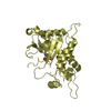
 Movie
Movie Controller


+ Open data

- Basic information
| Entry | Database: PDB / ID: 2ooq | ||||||
|---|---|---|---|---|---|---|---|
| Title | Crystal Structure of the Human Receptor Phosphatase PTPRT | ||||||
 Components Components | Receptor-type tyrosine-protein phosphatase T | ||||||
 Keywords Keywords |  HYDROLASE / Protein Tyrosine Phosphatase / receptor / Human / Structural Genomics / Structural Genomics Consortium / SGC HYDROLASE / Protein Tyrosine Phosphatase / receptor / Human / Structural Genomics / Structural Genomics Consortium / SGC | ||||||
| Function / homology |  Function and homology informationtransmembrane receptor protein tyrosine phosphatase activity / gamma-catenin binding / delta-catenin binding / alpha-catenin binding / cellular response to interleukin-6 / negative regulation of receptor signaling pathway via STAT / STAT family protein binding / peptidyl-tyrosine dephosphorylation involved in inactivation of protein kinase activity / homophilic cell adhesion via plasma membrane adhesion molecules / plasma membrane => GO:0005886 ...transmembrane receptor protein tyrosine phosphatase activity / gamma-catenin binding / delta-catenin binding / alpha-catenin binding / cellular response to interleukin-6 / negative regulation of receptor signaling pathway via STAT / STAT family protein binding / peptidyl-tyrosine dephosphorylation involved in inactivation of protein kinase activity / homophilic cell adhesion via plasma membrane adhesion molecules / plasma membrane => GO:0005886 / peptidyl-tyrosine dephosphorylation / protein dephosphorylation / protein-tyrosine-phosphatase / negative regulation of cell migration / protein tyrosine phosphatase activity / beta-catenin binding / cell surface receptor protein tyrosine kinase signaling pathway / protein phosphatase binding / membrane => GO:0016020 / cell adhesion / cadherin binding / cell surface / signal transduction / protein homodimerization activity / plasma membrane Function and homology informationtransmembrane receptor protein tyrosine phosphatase activity / gamma-catenin binding / delta-catenin binding / alpha-catenin binding / cellular response to interleukin-6 / negative regulation of receptor signaling pathway via STAT / STAT family protein binding / peptidyl-tyrosine dephosphorylation involved in inactivation of protein kinase activity / homophilic cell adhesion via plasma membrane adhesion molecules / plasma membrane => GO:0005886 ...transmembrane receptor protein tyrosine phosphatase activity / gamma-catenin binding / delta-catenin binding / alpha-catenin binding / cellular response to interleukin-6 / negative regulation of receptor signaling pathway via STAT / STAT family protein binding / peptidyl-tyrosine dephosphorylation involved in inactivation of protein kinase activity / homophilic cell adhesion via plasma membrane adhesion molecules / plasma membrane => GO:0005886 / peptidyl-tyrosine dephosphorylation / protein dephosphorylation / protein-tyrosine-phosphatase / negative regulation of cell migration / protein tyrosine phosphatase activity / beta-catenin binding / cell surface receptor protein tyrosine kinase signaling pathway / protein phosphatase binding / membrane => GO:0016020 / cell adhesion / cadherin binding / cell surface / signal transduction / protein homodimerization activity / plasma membraneSimilarity search - Function | ||||||
| Biological species |  Homo sapiens (human) Homo sapiens (human) | ||||||
| Method | X-RAY DIFFRACTION / MOLECULAR REPLACEMENT / Resolution: 1.8 Å | ||||||
 Authors Authors | Ugochukwu, E. / Alfano, I. / Barr, A. / Keates, T. / Eswaran, J. / Salah, E. / Savitsky, P. / Bunkoczi, G. / Edwards, A. / Arrowsmith, C.H. ...Ugochukwu, E. / Alfano, I. / Barr, A. / Keates, T. / Eswaran, J. / Salah, E. / Savitsky, P. / Bunkoczi, G. / Edwards, A. / Arrowsmith, C.H. / Weigelt, J. / Sundstrom, M. / von Delft, F. / Knapp, S. / Structural Genomics Consortium (SGC) | ||||||
 Citation Citation | Journal: Cell(Cambridge,Mass.) / Year: 2009 Title: Large-scale structural analysis of the classical human protein tyrosine phosphatome. Authors: Barr, A.J. / Ugochukwu, E. / Lee, W.H. / King, O.N. / Filippakopoulos, P. / Alfano, I. / Savitsky, P. / Burgess-Brown, N.A. / Muller, S. / Knapp, S. | ||||||
| History |
|
- Structure visualization
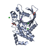
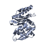
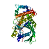
| Structure viewer | Molecule: MolmilJmol/JSmol |
|---|
- Downloads & links
-Download
| PDBx/mmCIF format | 2ooq.cif.gz | 137.9 KB | Display | PDBx/mmCIF format |
|---|---|---|---|---|
| PDB format | pdb2ooq.ent.gz | 105.1 KB | Display | PDB format |
| PDBx/mmJSON format | 2ooq.json.gz | Tree view | PDBx/mmJSON format | |
| Others |  Other downloads Other downloads |
-Validation report
| Arichive directory | https://data.pdbj.org/pub/pdb/validation_reports/oo/2ooqftp://data.pdbj.org/pub/pdb/validation_reports/oo/2ooq | HTTPS FTP |
|---|
-Related structure data
| Related structure data |  2ahsC  2b49C  2cfvC 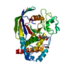 2cjzC  2gjtC  2h4vC  2i75C  2jjdC  2nlkC  2nz6C  2oc3C  2p6xC  2pa5C  2qepC  3b7oC 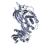 1larS  1rpmS  2c7sS 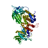 2fh7S S: Starting model for refinement C: citing same article ( |
|---|---|
| Similar structure data |


-Links
 PDBj
PDBj


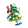
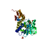
- Assembly
| Deposited unit | 
| |||||||||||||||||||||||||||||||||||||||||||||||||||||||||||||||||||||||||||||||||||||||||||
|---|---|---|---|---|---|---|---|---|---|---|---|---|---|---|---|---|---|---|---|---|---|---|---|---|---|---|---|---|---|---|---|---|---|---|---|---|---|---|---|---|---|---|---|---|---|---|---|---|---|---|---|---|---|---|---|---|---|---|---|---|---|---|---|---|---|---|---|---|---|---|---|---|---|---|---|---|---|---|---|---|---|---|---|---|---|---|---|---|---|---|---|---|
| 1 | 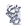
| |||||||||||||||||||||||||||||||||||||||||||||||||||||||||||||||||||||||||||||||||||||||||||
| 2 | 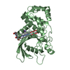
| |||||||||||||||||||||||||||||||||||||||||||||||||||||||||||||||||||||||||||||||||||||||||||
| Unit cell |
| |||||||||||||||||||||||||||||||||||||||||||||||||||||||||||||||||||||||||||||||||||||||||||
| Noncrystallographic symmetry (NCS) | NCS domain:
NCS domain segments: Ens-ID: 1 / Refine code: 5
|
-Components
| #1: Protein | Mass: 32763.082 Da / Num. of mol.: 2 / Fragment: PTPRT Source method: isolated from a genetically manipulated source Source: (gene. exp.) Homo sapiens (human) / Gene: PTPRT, KIAA0283 / Plasmid: pNIC28-Bsa4 / Production host:  Escherichia coli (E. coli) / Strain (production host): BL21 Rosetta (DE3) / References: UniProt: O14522, protein-tyrosine-phosphatase Escherichia coli (E. coli) / Strain (production host): BL21 Rosetta (DE3) / References: UniProt: O14522, protein-tyrosine-phosphatase#2: Chemical | Bis-tris propane  Mass: 282.334 Da / Num. of mol.: 2 / Source method: obtained synthetically / Formula: C11H26N2O6 / Comment: pH buffer*YM Mass: 282.334 Da / Num. of mol.: 2 / Source method: obtained synthetically / Formula: C11H26N2O6 / Comment: pH buffer*YM#3: Chemical | ChemComp-NA / |   Mass: 22.990 Da / Num. of mol.: 1 / Source method: obtained synthetically / Formula: Na Mass: 22.990 Da / Num. of mol.: 1 / Source method: obtained synthetically / Formula: Na#4: Chemical | ChemComp-EDO / | Ethylene glycol  Mass: 62.068 Da / Num. of mol.: 1 / Source method: obtained synthetically / Formula: C2H6O2 Mass: 62.068 Da / Num. of mol.: 1 / Source method: obtained synthetically / Formula: C2H6O2#5: Water | ChemComp-HOH / | Water Mass: 18.015 Da / Num. of mol.: 436 / Source method: isolated from a natural source / Formula: H2O Mass: 18.015 Da / Num. of mol.: 436 / Source method: isolated from a natural source / Formula: H2O |
|---|
-Experimental details
-Experiment
| Experiment | Method: X-RAY DIFFRACTION / Number of used crystals: 1 |
|---|
- Sample preparation
| Crystal | Density Matthews: 2.21 Å3/Da / Density % sol: 44.28 % |
|---|---|
| Crystal grow | Temperature: 293 K / Method: vapor diffusion, sitting drop / pH: 7 Details: 0.2M LiCl, 0.1M HEPES, 20.0% PEG 6000, 10.0% Ethylene glycol, pH 7.0, VAPOR DIFFUSION, SITTING DROP, temperature 293K |
-Data collection
| Diffraction | Mean temperature: 100 K |
|---|---|
| Diffraction source | Source: ROTATING ANODE / Type: RIGAKU RU200 / Wavelength: 1.5418 Å |
| Detector | Type: RIGAKU RAXIS HTC / Detector: IMAGE PLATE / Date: Jan 7, 2007 |
| Radiation | Protocol: SINGLE WAVELENGTH / Monochromatic (M) / Laue (L): M / Scattering type: x-ray |
| Radiation wavelength | Wavelength: 1.5418 Å / Relative weight: 1 |
| Reflection | Resolution: 1.8→89.77 Å / Num. obs: 52802 / % possible obs: 100 % / Observed criterion σ(F): 0 / Observed criterion σ(I): 0 / Redundancy: 3.7 % / Biso Wilson estimate: 18.6 Å2 / Rmerge(I) obs: 0.092 / Rsym value: 0.092 / Net I/σ(I): 16 |
| Reflection shell | Resolution: 1.8→1.9 Å / Redundancy: 3.6 % / Rmerge(I) obs: 0.595 / Mean I/σ(I) obs: 2.6 / Rsym value: 0.595 / % possible all: 100 |
- Processing
| Software |
| |||||||||||||||||||||||||||||||||||||||||||||||||||||||||||||||||||||||||||||||||||||||||||||||||||||||||||||||||||||||||||||
|---|---|---|---|---|---|---|---|---|---|---|---|---|---|---|---|---|---|---|---|---|---|---|---|---|---|---|---|---|---|---|---|---|---|---|---|---|---|---|---|---|---|---|---|---|---|---|---|---|---|---|---|---|---|---|---|---|---|---|---|---|---|---|---|---|---|---|---|---|---|---|---|---|---|---|---|---|---|---|---|---|---|---|---|---|---|---|---|---|---|---|---|---|---|---|---|---|---|---|---|---|---|---|---|---|---|---|---|---|---|---|---|---|---|---|---|---|---|---|---|---|---|---|---|---|---|---|
| Refinement | Method to determine structure: MOLECULAR REPLACEMENT Starting model: PDB entries 1RPM, 2C7S, 1LAR, 2FH7 Resolution: 1.8→89.77 Å / Cor.coef. Fo:Fc: 0.968 / Cor.coef. Fo:Fc free: 0.951 / SU B: 4.669 / SU ML: 0.075 / TLS residual ADP flag: LIKELY RESIDUAL / Cross valid method: THROUGHOUT / σ(F): 0 / ESU R: 0.107 / ESU R Free: 0.108 / Stereochemistry target values: MAXIMUM LIKELIHOOD / Details: HYDROGENS HAVE BEEN ADDED IN THE RIDING POSITIONS
| |||||||||||||||||||||||||||||||||||||||||||||||||||||||||||||||||||||||||||||||||||||||||||||||||||||||||||||||||||||||||||||
| Solvent computation | Ion probe radii: 0.8 Å / Shrinkage radii: 0.8 Å / VDW probe radii: 1.4 Å / Solvent model: BABINET MODEL WITH MASK | |||||||||||||||||||||||||||||||||||||||||||||||||||||||||||||||||||||||||||||||||||||||||||||||||||||||||||||||||||||||||||||
| Displacement parameters | Biso mean: 22.417 Å2
| |||||||||||||||||||||||||||||||||||||||||||||||||||||||||||||||||||||||||||||||||||||||||||||||||||||||||||||||||||||||||||||
| Refinement step | Cycle: LAST / Resolution: 1.8→89.77 Å
| |||||||||||||||||||||||||||||||||||||||||||||||||||||||||||||||||||||||||||||||||||||||||||||||||||||||||||||||||||||||||||||
| Refine LS restraints |
| |||||||||||||||||||||||||||||||||||||||||||||||||||||||||||||||||||||||||||||||||||||||||||||||||||||||||||||||||||||||||||||
| Refine LS restraints NCS | Dom-ID: 1 / Auth asym-ID: A / Ens-ID: 1 / Refine-ID: X-RAY DIFFRACTION
| |||||||||||||||||||||||||||||||||||||||||||||||||||||||||||||||||||||||||||||||||||||||||||||||||||||||||||||||||||||||||||||
| LS refinement shell | Resolution: 1.8→1.85 Å / Total num. of bins used: 20
| |||||||||||||||||||||||||||||||||||||||||||||||||||||||||||||||||||||||||||||||||||||||||||||||||||||||||||||||||||||||||||||
| Refinement TLS params. | Method: refined / Refine-ID: X-RAY DIFFRACTION
| |||||||||||||||||||||||||||||||||||||||||||||||||||||||||||||||||||||||||||||||||||||||||||||||||||||||||||||||||||||||||||||
| Refinement TLS group |
|
