 Movie
Movie Controller

[English] 日本語
 Yorodumi
- PDB-2oko: Z. mobilis tRNA guanine transglycosylase E235Q mutant apo-structu... -
+ Open data

- Basic information
| Entry | Database: PDB / ID: 2oko | ||||||
|---|---|---|---|---|---|---|---|
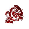
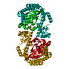
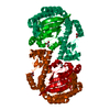
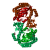
| Title | Z. mobilis tRNA guanine transglycosylase E235Q mutant apo-structure at pH 5.5 | ||||||
 Components Components | Queuine tRNA-ribosyltransferase | ||||||
 Keywords Keywords | TRANSFERASE / tRNA guanine transglycosylase / TGT / PreQ0 / E235Q mutation | ||||||
| Function / homology |  Function and homology information Function and homology informationtRNA-guanosine34 preQ1 transglycosylase / tRNA wobble guanine modification / tRNA-guanosine(34) queuine transglycosylase activity / tRNA-guanine transglycosylation / queuosine biosynthetic process / metal ion binding / cytosolSimilarity search - Function | ||||||
| Biological species |  Zymomonas mobilis (bacteria) Zymomonas mobilis (bacteria) | ||||||
| Method | X-RAY DIFFRACTION / MOLECULAR REPLACEMENT / Resolution: 1.5 Å | ||||||
 Authors Authors | Tidten, N. | ||||||
 Citation Citation | Journal: J.Mol.Biol. / Year: 2007 Title: Glutamate versus Glutamine Exchange Swaps Substrate Selectivity in tRNA-Guanine Transglycosylase: Insight into the Regulation of Substrate Selectivity by Kinetic and Crystallographic Studies. Authors: Tidten, N. / Stengl, B. / Heine, A. / Garcia, G.A. / Klebe, G. / Reuter, K. | ||||||
| History |
| ||||||
| Remark 999 | SEQUENCE THE CONFLICT INVOLVING RESIDUE 311 IS CONSISTENT WITH THE REF. 1 AND 2 IN THE UNIPROT ...SEQUENCE THE CONFLICT INVOLVING RESIDUE 311 IS CONSISTENT WITH THE REF. 1 AND 2 IN THE UNIPROT SEQUENCE DATABASE, TGT_ZYMMO |
- Structure visualization
| Structure viewer | Molecule: MolmilJmol/JSmol |
|---|
- Downloads & links
-Download
| PDBx/mmCIF format | 2oko.cif.gz | 178.1 KB | Display | PDBx/mmCIF format |
|---|---|---|---|---|
| PDB format | pdb2oko.ent.gz | 139.9 KB | Display | PDB format |
| PDBx/mmJSON format | 2oko.json.gz | Tree view | PDBx/mmJSON format | |
| Others |  Other downloads Other downloads |
-Validation report
| Arichive directory | https://data.pdbj.org/pub/pdb/validation_reports/ok/2okoftp://data.pdbj.org/pub/pdb/validation_reports/ok/2oko | HTTPS FTP |
|---|
-Related structure data
| Related structure data |  2potC  2pwuC  2pwvC  2qiiC 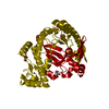 2z1vC 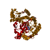 2z1wC 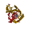 2z1xC  1y5xS S: Starting model for refinement C: citing same article ( |
|---|---|
| Similar structure data |


-Links
 PDBj
PDBj- Assembly
| Deposited unit | 
| ||||||||
|---|---|---|---|---|---|---|---|---|---|
| 1 | 
| ||||||||
| Unit cell |
| ||||||||
| Components on special symmetry positions |
| ||||||||
| Details | The second part of the biological assembly is generated by the two fold axis |
-Components
| #1: Protein | / tRNA-guanine transglycosylase / Guanine insertion enzyme Mass: 42793.523 Da / Num. of mol.: 1 / Mutation: E235Q Source method: isolated from a genetically manipulated source Source: (gene. exp.) Zymomonas mobilis (bacteria) / Gene: tgt / Plasmid: pET9d / Production host: Escherichia coli (E. coli) / Strain (production host): BL21(DE3)pLysSReferences: UniProt: P28720, tRNA-guanosine34 preQ1 transglycosylase | ||
|---|---|---|---|
| #2: Chemical | ChemComp-ZN /   Mass: 65.409 Da / Num. of mol.: 1 / Source method: obtained synthetically / Formula: Zn Mass: 65.409 Da / Num. of mol.: 1 / Source method: obtained synthetically / Formula: Zn | ||
| #3: Chemical | ChemComp-GOL / Glycerol  Mass: 92.094 Da / Num. of mol.: 16 / Source method: obtained synthetically / Formula: C3H8O3 Mass: 92.094 Da / Num. of mol.: 16 / Source method: obtained synthetically / Formula: C3H8O3#4: Water | ChemComp-HOH / | Water Mass: 18.015 Da / Num. of mol.: 346 / Source method: isolated from a natural source / Formula: H2O Mass: 18.015 Da / Num. of mol.: 346 / Source method: isolated from a natural source / Formula: H2O |
-Experimental details
-Experiment
| Experiment | Method: X-RAY DIFFRACTION |
|---|
- Sample preparation
| Crystal | Density Matthews: 2.33 Å3/Da / Density % sol: 49.01 % |
|---|---|
| Crystal grow | pH: 5.5 / Details: pH 5.5 |
-Data collection
| Diffraction source | Source: ROTATING ANODE / Type: RIGAKU / Wavelength: 1.5418 |
|---|---|
| Detector | Type: RIGAKU RAXIS IV / Detector: IMAGE PLATE / Date: Dec 13, 2006 |
| Radiation | Protocol: SINGLE WAVELENGTH / Monochromatic (M) / Laue (L): M / Scattering type: x-ray |
| Radiation wavelength | Wavelength: 1.5418 Å / Relative weight: 1 |
| Reflection | Resolution: 1.5→50 Å / Num. obs: 210241 / % possible obs: 91.5 % / Redundancy: 3.5 % / Rmerge(I) obs: 0.1224 / Rsym value: 0.032 / Net I/σ(I): 33 |
- Processing
| Software |
| |||||||||||||||||||||||||||||||||
|---|---|---|---|---|---|---|---|---|---|---|---|---|---|---|---|---|---|---|---|---|---|---|---|---|---|---|---|---|---|---|---|---|---|---|
| Refinement | Method to determine structure: MOLECULAR REPLACEMENT Starting model: PDB ENTRY 1Y5X Resolution: 1.5→10 Å / Num. parameters: 31053 / Num. restraintsaints: 38625 / Cross valid method: FREE R / σ(F): 0 / Stereochemistry target values: ENGH AND HUBER Details: ANISOTROPIC SCALING APPLIED BY THE METHOD OF PARKIN, MOEZZI & HOPE, J.APPL.CRYST.28(1995)53-56 ANISOTROPIC REFINEMENT REDUCED FREE R (NO CUTOFF) BY ?
| |||||||||||||||||||||||||||||||||
| Solvent computation | Solvent model: BABINET (SWAT) | |||||||||||||||||||||||||||||||||
| Refine analyze | Num. disordered residues: 19 / Occupancy sum hydrogen: 2810 / Occupancy sum non hydrogen: 3355.5 | |||||||||||||||||||||||||||||||||
| Refinement step | Cycle: LAST / Resolution: 1.5→10 Å
| |||||||||||||||||||||||||||||||||
| Refine LS restraints |
|
