 Movie
Movie Controller

[English] 日本語
 Yorodumi
- PDB-2ojx: Molecular and structural basis of polo-like kinase 1 substrate re... -
+ Open data

- Basic information
| Entry | Database: PDB / ID: 2ojx | ||||||
|---|---|---|---|---|---|---|---|
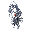
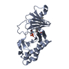
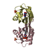
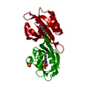
| Title | Molecular and structural basis of polo-like kinase 1 substrate recognition: Implications in centrosomal localization | ||||||
 Components Components |
| ||||||
 Keywords Keywords |  TRANSFERASE / POLO BOX DOMAIN / KINASE / centrosome TRANSFERASE / POLO BOX DOMAIN / KINASE / centrosome | ||||||
| Function / homology |  Function and homology information Function and homology informationpositive regulation of G2/MI transition of meiotic cell cycle / Mitotic Telophase/Cytokinesis / regulation of protein localization to cell cortex / Mitotic Metaphase/Anaphase Transition / Golgi inheritance / synaptonemal complex disassembly / Activation of NIMA Kinases NEK9, NEK6, NEK7 / homologous chromosome segregation / polo kinase / nuclear membrane disassembly ...positive regulation of G2/MI transition of meiotic cell cycle / Mitotic Telophase/Cytokinesis / regulation of protein localization to cell cortex / Mitotic Metaphase/Anaphase Transition / Golgi inheritance / synaptonemal complex disassembly / Activation of NIMA Kinases NEK9, NEK6, NEK7 / homologous chromosome segregation / polo kinase / nuclear membrane disassembly / mitotic nuclear membrane disassembly / protein localization to nuclear envelope / Phosphorylation of Emi1 / WW domain binding / metaphase/anaphase transition of mitotic cell cycle / synaptonemal complex / female meiosis chromosome segregation / Phosphorylation of the APC/C / regulation of protein binding / anaphase-promoting complex binding / outer kinetochore / negative regulation of cyclin-dependent protein serine/threonine kinase activity / positive regulation of ubiquitin protein ligase activity / regulation of mitotic spindle assembly / microtubule bundle formation / Polo-like kinase mediated events / Golgi Cisternae Pericentriolar Stack Reorganization / mitotic chromosome condensation / regulation of mitotic metaphase/anaphase transition / sister chromatid cohesion / positive regulation of ubiquitin-protein transferase activity / centrosome cycle / double-strand break repair via alternative nonhomologous end joining / TP53 regulates transcription of additional cell cycle genes whose exact role in the p53 pathway remain uncertain / regulation of mitotic cell cycle phase transition / regulation of mitotic nuclear division / Deregulated CDK5 triggers multiple neurodegenerative pathways in Alzheimer's disease models / mitotic spindle assembly checkpoint signaling / mitotic spindle pole / regulation of cyclin-dependent protein serine/threonine kinase activity / regulation of anaphase-promoting complex-dependent catabolic process / mitotic G2 DNA damage checkpoint signaling / mitotic sister chromatid segregation / establishment of mitotic spindle orientation / phosphoprotein phosphatase activity / positive regulation of proteolysis / mitotic cytokinesis / centriolar satellite / spindle midzone / negative regulation of double-strand break repair via homologous recombination / Amplification of signal from unattached kinetochores via a MAD2 inhibitory signal / Chk1/Chk2(Cds1) mediated inactivation of Cyclin B:Cdk1 complex / Activation of ATR in response to replication stress / Cyclin A/B1/B2 associated events during G2/M transition / Mitotic Prometaphase / EML4 and NUDC in mitotic spindle formation / RHO GTPases activate PKNs / Loss of Nlp from mitotic centrosomes / Loss of proteins required for interphase microtubule organization from the centrosome / Recruitment of mitotic centrosome proteins and complexes / protein localization to chromatin / Resolution of Sister Chromatid Cohesion / Recruitment of NuMA to mitotic centrosomes / Anchoring of the basal body to the plasma membrane / regulation of mitotic cell cycle / positive regulation of G2/M transition of mitotic cell cycle / centriole / AURKA Activation by TPX2 / protein-tyrosine-phosphatase / TP53 Regulates Transcription of Genes Involved in G2 Cell Cycle Arrest / Condensation of Prophase Chromosomes / mitotic spindle organization / positive regulation of peptidyl-threonine phosphorylation / regulation of cytokinesis / protein tyrosine phosphatase activity / RHO GTPases Activate Formins / protein destabilization / APC/C:Cdh1 mediated degradation of Cdc20 and other APC/C:Cdh1 targeted proteins in late mitosis/early G1 / establishment of protein localization / mitochondrial intermembrane space / kinetochore / spindle pole / spindle / positive regulation of protein localization to nucleus / Separation of Sister Chromatids / The role of GTSE1 in G2/M progression after G2 checkpoint / G2/M transition of mitotic cell cycle / microtubule cytoskeleton / Regulation of PLK1 Activity at G2/M Transition / double-strand break repair / positive regulation of proteasomal ubiquitin-dependent protein catabolic process / mitotic cell cycle / midbody / microtubule binding / spermatogenesis / peptidyl-serine phosphorylation / cell population proliferation / protein ubiquitination / regulation of cell cycle / protein kinase activitySimilarity search - Function | ||||||
| Biological species |  Homo sapiens (human) Homo sapiens (human) | ||||||
| Method | X-RAY DIFFRACTION / SYNCHROTRON / MOLECULAR REPLACEMENT / Resolution: 2.85 Å | ||||||
 Authors Authors | Garcia-Alvarez, B. / de Carcer, G. / Ibanez, S. / Bragado-Nilsson, E. / Montoya, G. | ||||||
 Citation Citation | Journal: Proc.Natl.Acad.Sci.Usa / Year: 2007 Title: Molecular and structural basis of polo-like kinase 1 substrate recognition: Implications in centrosomal localization. Authors: Garcia-Alvarez, B. / de Carcer, G. / Ibanez, S. / Bragado-Nilsson, E. / Montoya, G. | ||||||
| History |
|
- Structure visualization
| Structure viewer | Molecule: MolmilJmol/JSmol |
|---|
- Downloads & links
-Download
| PDBx/mmCIF format | 2ojx.cif.gz | 56.3 KB | Display | PDBx/mmCIF format |
|---|---|---|---|---|
| PDB format | pdb2ojx.ent.gz | 40 KB | Display | PDB format |
| PDBx/mmJSON format | 2ojx.json.gz | Tree view | PDBx/mmJSON format | |
| Others |  Other downloads Other downloads |
-Validation report
| Arichive directory | https://data.pdbj.org/pub/pdb/validation_reports/oj/2ojxftp://data.pdbj.org/pub/pdb/validation_reports/oj/2ojx | HTTPS FTP |
|---|
-Related structure data
| Related structure data |  2ogqC  3bziC  1umwS C: citing same article ( S: Starting model for refinement |
|---|---|
| Similar structure data |


-Links
 PDBj
PDBj


- Assembly
| Deposited unit | 
| ||||||||
|---|---|---|---|---|---|---|---|---|---|
| 1 |
| ||||||||
| Unit cell |
|
-Components
| #1: Protein | Mass: 27502.334 Da / Num. of mol.: 1 / Fragment: residues 365-603 Source method: isolated from a genetically manipulated source Source: (gene. exp.) Homo sapiens (human) / Gene: PLK1, PLK / Plasmid: PGEX-6P-2 / Production host:  Escherichia coli (E. coli) / Strain (production host): BL21-pLys / References: UniProt: P53350, polo kinase Escherichia coli (E. coli) / Strain (production host): BL21-pLys / References: UniProt: P53350, polo kinase |
|---|---|
| #2: Protein/peptide | Peptide synthesis Mass: 917.082 Da / Num. of mol.: 1 / Source method: obtained synthetically / Details: Synthetic peptide / References: UniProt: P30307*PLUS |
| #3: Water | ChemComp-HOH / Water Mass: 18.015 Da / Num. of mol.: 10 / Source method: isolated from a natural source / Formula: H2O Mass: 18.015 Da / Num. of mol.: 10 / Source method: isolated from a natural source / Formula: H2O |
-Experimental details
-Experiment
| Experiment | Method: X-RAY DIFFRACTION / Number of used crystals: 1 |
|---|
- Sample preparation
| Crystal | Density Matthews: 1.84 Å3/Da / Density % sol: 33.25 % |
|---|---|
| Crystal grow | Temperature: 289 K / Method: vapor diffusion, hanging drop / pH: 7.5 Details: 20 % PEG 10000, pH 7.5, VAPOR DIFFUSION, HANGING DROP, temperature 289K |
-Data collection
| Diffraction | Mean temperature: 100 K |
|---|---|
| Diffraction source | Source: SYNCHROTRON / Site: SLS  / Beamline: X06SA / Beamline: X06SA |
| Detector | Type: MARMOSAIC 225 mm CCD / Detector: CCD / Date: Jul 1, 2005 |
| Radiation | Protocol: SINGLE WAVELENGTH / Monochromatic (M) / Laue (L): M / Scattering type: x-ray |
| Radiation wavelength | Relative weight: 1 |
| Reflection | Resolution: 2.85→50 Å / Num. obs: 4971 / % possible obs: 96.3 % / Observed criterion σ(I): 1 / Redundancy: 2.6 % / Rsym value: 0.09 / Net I/σ(I): 4.8 |
| Reflection shell | Resolution: 2.85→3 Å / Redundancy: 2.6 % / Mean I/σ(I) obs: 1.9 / Num. unique all: 618 / Rsym value: 0.31 / % possible all: 96.3 |
- Processing
| Software |
| ||||||||||||||||
|---|---|---|---|---|---|---|---|---|---|---|---|---|---|---|---|---|---|
| Refinement | Method to determine structure: MOLECULAR REPLACEMENT Starting model: PDB ENTRY 1UMW Resolution: 2.85→18.94 Å / Isotropic thermal model: Isotropic / Cross valid method: THROUGHOUT / σ(I): 1
| ||||||||||||||||
| Refinement step | Cycle: LAST / Resolution: 2.85→18.94 Å
|
