Type: MARRESEARCH / Detector: CCD / Date: May 18, 2006 / Details: BENT MIRROR
Radiation
Monochromator: TRIANGULAR MONOCHROMATOR / Protocol: SINGLE WAVELENGTH / Monochromatic (M) / Laue (L): M / Scattering type: x-ray
Radiation wavelength
Wavelength: 1.05 Å / Relative weight: 1
Reflection
Resolution: 1.5→20 Å / Num. obs: 75246 / % possible obs: 91.2 % / Redundancy: 3.1 % / Biso Wilson estimate: 19.8 Å2 / Rsym value: 0.06 / Net I/σ(I): 30.8
Reflection shell
Resolution: 1.5→1.52 Å / Redundancy: 2.6 % / Mean I/σ(I) obs: 4.1 / Rsym value: 0.23 / % possible all: 99.6
-
Processing
Software
Name
Version
Classification
SHELXD
phasing
SHARP
phasing
NCSREF
modelbuilding
REFMAC
5.1.24
refinement
DENZO
datareduction
SCALEPACK
datascaling
DM
phasing
ARP/wARP
modelbuilding
Refinement
Method to determine structure: SAD / Resolution: 1.5→19.8 Å / Cor.coef. Fo:Fc: 0.967 / Cor.coef. Fo:Fc free: 0.955 / SU B: 1.32 / SU ML: 0.05 / Cross valid method: THROUGHOUT / ESU R: 0.08 / ESU R Free: 0.08 / Stereochemistry target values: MAXIMUM LIKELIHOOD Details: MISSING RESOLUTION SHELL DUE TO THE PRESENCE OF AN ICE RING. IN THE CRYSTALLIZATION BUFFER CD2+, CA2+ AND NA+ IONS WERE PRESENT. ALL METAL CATIONS IN THE DENSITY WERE MODELLED AS CD2+ IONS, ...Details: MISSING RESOLUTION SHELL DUE TO THE PRESENCE OF AN ICE RING. IN THE CRYSTALLIZATION BUFFER CD2+, CA2+ AND NA+ IONS WERE PRESENT. ALL METAL CATIONS IN THE DENSITY WERE MODELLED AS CD2+ IONS, BUT IN SOME CASES THEIR IDENTITY IS UNCERTAIN. THE ACTIVE SITE IS DISTORTED AND THE METAL IONS ARE NOT BOUND TO THE ACTIVE SITE. TLS REFINEMENT USED. HYDROGENS HAVE BEEN ADDED IN THE RIDING POSITIONS. BOTH REFMAC AND CNS ARE USED FOR REFINEMENT.
Rfactor
Num. reflection
% reflection
Selection details
Rfree
0.212
3629
5 %
RANDOM
Rwork
0.184
-
-
-
obs
0.186
72535
93.9 %
-
all
-
72535
-
-
Solvent computation
Ion probe radii: 0.8 Å / Shrinkage radii: 0.8 Å / VDW probe radii: 1.4 Å / Solvent model: BABINET MODEL WITH MASK
Displacement parameters
Biso mean: 17.4 Å2
Baniso -1
Baniso -2
Baniso -3
1-
1.06 Å2
0 Å2
-0.44 Å2
2-
-
0.86 Å2
0 Å2
3-
-
-
-2.09 Å2
Refinement step
Cycle: LAST / Resolution: 1.5→19.8 Å
Protein
Nucleic acid
Ligand
Solvent
Total
Num. atoms
3847
0
25
288
4160
Refine LS restraints
Refine-ID
Type
Dev ideal
Dev ideal target
Number
X-RAY DIFFRACTION
r_bond_refined_d
0.014
0.021
3858
X-RAY DIFFRACTION
r_bond_other_d
0.007
0.02
3380
X-RAY DIFFRACTION
r_angle_refined_deg
1.525
1.946
5205
X-RAY DIFFRACTION
r_angle_other_deg
0.821
3
7899
X-RAY DIFFRACTION
r_dihedral_angle_1_deg
6.63
5
472
X-RAY DIFFRACTION
r_dihedral_angle_2_deg
X-RAY DIFFRACTION
r_dihedral_angle_3_deg
X-RAY DIFFRACTION
r_dihedral_angle_4_deg
X-RAY DIFFRACTION
r_chiral_restr
0.1
0.2
574
X-RAY DIFFRACTION
r_gen_planes_refined
0.008
0.02
4315
X-RAY DIFFRACTION
r_gen_planes_other
0.01
0.02
750
X-RAY DIFFRACTION
r_nbd_refined
0.227
0.2
716
X-RAY DIFFRACTION
r_nbd_other
0.251
0.2
3895
X-RAY DIFFRACTION
r_nbtor_refined
X-RAY DIFFRACTION
r_nbtor_other
0.085
0.2
2165
X-RAY DIFFRACTION
r_xyhbond_nbd_refined
0.189
0.2
216
X-RAY DIFFRACTION
r_xyhbond_nbd_other
X-RAY DIFFRACTION
r_metal_ion_refined
0.211
X-RAY DIFFRACTION
r_metal_ion_other
X-RAY DIFFRACTION
r_symmetry_vdw_refined
0.239
0.2
21
X-RAY DIFFRACTION
r_symmetry_vdw_other
0.336
0.2
89
X-RAY DIFFRACTION
r_symmetry_hbond_refined
0.233
0.2
18
X-RAY DIFFRACTION
r_symmetry_hbond_other
X-RAY DIFFRACTION
r_symmetry_metal_ion_refined
X-RAY DIFFRACTION
r_symmetry_metal_ion_other
X-RAY DIFFRACTION
r_mcbond_it
1.012
1.5
2351
X-RAY DIFFRACTION
r_mcbond_other
X-RAY DIFFRACTION
r_mcangle_it
1.801
2
3778
X-RAY DIFFRACTION
r_scbond_it
2.66
3
1507
X-RAY DIFFRACTION
r_scangle_it
3.979
4.5
1427
X-RAY DIFFRACTION
r_rigid_bond_restr
X-RAY DIFFRACTION
r_sphericity_free
X-RAY DIFFRACTION
r_sphericity_bonded
LS refinement shell
Resolution: 1.5→1.54 Å / Total num. of bins used: 20 /
Rfactor
Num. reflection
Rfree
0.32
268
Rwork
0.29
5727
Refinement TLS params.
Method: refined / Refine-ID: X-RAY DIFFRACTION
ID
L11 (°2)
L12 (°2)
L13 (°2)
L22 (°2)
L23 (°2)
L33 (°2)
S11 (Å °)
S12 (Å °)
S13 (Å °)
S21 (Å °)
S22 (Å °)
S23 (Å °)
S31 (Å °)
S32 (Å °)
S33 (Å °)
T11 (Å2)
T12 (Å2)
T13 (Å2)
T22 (Å2)
T23 (Å2)
T33 (Å2)
Origin x (Å)
Origin y (Å)
Origin z (Å)
1
1.292
-0.8074
0.392
0.8981
-0.3018
1.1763
-0.0772
-0.0518
0.0985
0.0524
0.1011
-0.1401
-0.0792
0.1198
-0.0239
0.0271
0.0037
0.0161
0.0086
-0.0174
0.0843
52.387
-3.902
45.17
2
0.2749
-0.0406
-0.1699
0.4455
-0.1055
1.6191
-0.015
-0.0031
0.0008
0.0106
-0.0028
-0.0204
0
0.0575
0.0178
0.0146
-0.0142
0.0187
0.0261
-0.0103
0.0661
45.319
3.714
14.049
3
0.9764
0.4878
1.0079
1.038
0.9905
1.7437
-0.0272
-0.0877
0.0022
-0.0674
-0.0114
0.1175
-0.0137
-0.1916
0.0386
0.0325
-0.0172
0.0165
0.0216
-0.0039
0.0815
22.438
-6.274
28.694
4
0.2175
-0.0453
-0.2223
0.4643
0.2018
1.5621
-0.0212
0.0199
-0.0032
-0.014
-0.0153
-0.0051
0.0149
-0.0461
0.0365
0.0141
0.0154
0.0141
0.0215
-0.0016
0.0765
30.212
5.434
57.804
Refinement TLS group
ID
Refine-ID
Refine TLS-ID
Auth asym-ID
Label asym-ID
Auth seq-ID
Label seq-ID
1
X-RAY DIFFRACTION
1
A
A
-2 - 65
1 - 68
2
X-RAY DIFFRACTION
1
A
A
161 - 190
164 - 193
3
X-RAY DIFFRACTION
1
A
A
232 - 246
235 - 249
4
X-RAY DIFFRACTION
2
A
A
66 - 160
69 - 163
5
X-RAY DIFFRACTION
2
A
A
191 - 231
194 - 234
6
X-RAY DIFFRACTION
3
B
B
4 - 65
7 - 68
7
X-RAY DIFFRACTION
3
B
B
161 - 190
164 - 193
8
X-RAY DIFFRACTION
3
B
B
232 - 246
235 - 249
9
X-RAY DIFFRACTION
4
B
B
66 - 160
69 - 163
10
X-RAY DIFFRACTION
4
B
B
191 - 231
194 - 234
+
About Yorodumi
-
News
-
Feb 9, 2022. New format data for meta-information of EMDB entries
New format data for meta-information of EMDB entries
Version 3 of the EMDB header file is now the official format.
The previous official version 1.9 will be removed from the archive.
In the structure databanks used in Yorodumi, some data are registered as the other names, "COVID-19 virus" and "2019-nCoV". Here are the details of the virus and the list of structure data.
Jan 31, 2019. EMDB accession codes are about to change! (news from PDBe EMDB page)
EMDB accession codes are about to change! (news from PDBe EMDB page)
The allocation of 4 digits for EMDB accession codes will soon come to an end. Whilst these codes will remain in use, new EMDB accession codes will include an additional digit and will expand incrementally as the available range of codes is exhausted. The current 4-digit format prefixed with “EMD-” (i.e. EMD-XXXX) will advance to a 5-digit format (i.e. EMD-XXXXX), and so on. It is currently estimated that the 4-digit codes will be depleted around Spring 2019, at which point the 5-digit format will come into force.
The EM Navigator/Yorodumi systems omit the EMD- prefix.
Related info.:Q: What is EMD? / ID/Accession-code notation in Yorodumi/EM Navigator
Yorodumi is a browser for structure data from EMDB, PDB, SASBDB, etc.
This page is also the successor to EM Navigator detail page, and also detail information page/front-end page for Omokage search.
The word "yorodu" (or yorozu) is an old Japanese word meaning "ten thousand". "mi" (miru) is to see.
Related info.:EMDB / PDB / SASBDB / Comparison of 3 databanks / Yorodumi Search / Aug 31, 2016. New EM Navigator & Yorodumi / Yorodumi Papers / Jmol/JSmol / Function and homology information / Changes in new EM Navigator and Yorodumi
 Movie
Movie Controller


 Open data
 Basic information
 Components
 Keywords
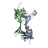
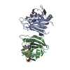
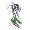
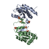
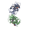
 HYDROLASE / MONOMERIC ENDONUCLEASE /
  Function and homology information
 Authors
 Citation
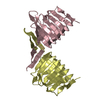
 Structure visualization
 Downloads & links
 Other downloads


 PDBj
PDBj
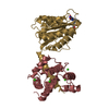
 Assembly


 Mass: 35.453 Da / Num. of mol.: 2 / Source method: obtained synthetically / Formula: Cl
 Mass: 112.411 Da / Num. of mol.: 15 / Source method: obtained synthetically / Formula: Cd
 Mass: 59.044 Da / Num. of mol.: 2 / Source method: obtained synthetically / Formula: C2H3O2
 Mass: 18.015 Da / Num. of mol.: 288 / Source method: isolated from a natural source / Formula: H2O
 Sample preparation
 / Beamline: BW6 / Wavelength: 1.05
  Processing