- PDB-1k8f: CRYSTAL STRUCTURE OF THE HUMAN C-TERMINAL CAP1-ADENYLYL CYCLASE A... -
+
Open data
ID or keywords:
Loading...
-
Basic information
Entry
Database: PDB / ID: 1k8f
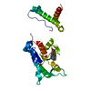
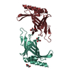
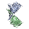
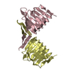
Title
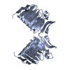
CRYSTAL STRUCTURE OF THE HUMAN C-TERMINAL CAP1-ADENYLYL CYCLASE ASSOCIATED PROTEIN
Components
ADENYLYL CYCLASE-ASSOCIATED PROTEIN
Keywords
UNKNOWN FUNCTION / CAP / CAP1 / ADENYLYL CYCLASE ASSOCIATED PROTEIN / ACTIN BINDING / Structural Genomics / PSI / Protein Structure Initiative / New York SGX Research Center for Structural Genomics / NYSGXRC
Function / homology
Function and homology information
regulation of adenylate cyclase activity / ameboidal-type cell migration / neutrophil degranulation / Role of ABL in ROBO-SLIT signaling / actin polymerization or depolymerization / establishment or maintenance of cell polarity / cortical actin cytoskeleton / adenylate cyclase binding / activation of adenylate cyclase activity / receptor-mediated endocytosis ...regulation of adenylate cyclase activity / ameboidal-type cell migration / neutrophil degranulation / Role of ABL in ROBO-SLIT signaling / actin polymerization or depolymerization / establishment or maintenance of cell polarity / cortical actin cytoskeleton / adenylate cyclase binding / activation of adenylate cyclase activity / receptor-mediated endocytosis / cell morphogenesis / azurophil granule lumen / Platelet degranulation / actin binding / focal adhesion / Neutrophil degranulation / signal transduction / extracellular exosome / extracellular region / plasma membrane Similarity search - Function
Adenylyl cyclase-associated protein CAP1 / Adenylate cyclase-associated CAP, N-terminal / CAP, conserved site, N-terminal / CAP, conserved site, C-terminal / Adenylate cyclase-associated CAP, N-terminal domain superfamily / Adenylate cyclase associated (CAP) N terminal / CAP protein signature 1. / CAP protein signature 2. / Adenylate cyclase-associated CAP / Adenylate cyclase-associated CAP, C-terminal ...Adenylyl cyclase-associated protein CAP1 / Adenylate cyclase-associated CAP, N-terminal / CAP, conserved site, N-terminal / CAP, conserved site, C-terminal / Adenylate cyclase-associated CAP, N-terminal domain superfamily / Adenylate cyclase associated (CAP) N terminal / CAP protein signature 1. / CAP protein signature 2. / Adenylate cyclase-associated CAP / Adenylate cyclase-associated CAP, C-terminal / Adenylate cyclase associated (CAP) C terminal / Pectate Lyase C-like - #70 / Adenylate cyclase-associated CAP, C-terminal superfamily / CARP motif / Domain in CAPs (cyclase-associated proteins) and X-linked retinitis pigmentosa 2 gene product. / C-CAP/cofactor C-like domain / C-CAP/cofactor C-like domain profile. / Cyclase-associated protein CAP/septum formation inhibitor MinC, C-terminal / Pectate Lyase C-like / 3 Solenoid / Mainly Beta Similarity search - Domain/homology
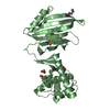
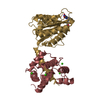
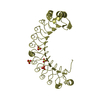
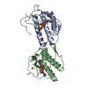
A: ADENYLYL CYCLASE-ASSOCIATED PROTEIN B: ADENYLYL CYCLASE-ASSOCIATED PROTEIN C: ADENYLYL CYCLASE-ASSOCIATED PROTEIN D: ADENYLYL CYCLASE-ASSOCIATED PROTEIN
In the structure databanks used in Yorodumi, some data are registered as the other names, "COVID-19 virus" and "2019-nCoV". Here are the details of the virus and the list of structure data.
Jan 31, 2019. EMDB accession codes are about to change! (news from PDBe EMDB page)
EMDB accession codes are about to change! (news from PDBe EMDB page)
The allocation of 4 digits for EMDB accession codes will soon come to an end. Whilst these codes will remain in use, new EMDB accession codes will include an additional digit and will expand incrementally as the available range of codes is exhausted. The current 4-digit format prefixed with “EMD-” (i.e. EMD-XXXX) will advance to a 5-digit format (i.e. EMD-XXXXX), and so on. It is currently estimated that the 4-digit codes will be depleted around Spring 2019, at which point the 5-digit format will come into force.
The EM Navigator/Yorodumi systems omit the EMD- prefix.
Related info.:Q: What is EMD? / ID/Accession-code notation in Yorodumi/EM Navigator
Yorodumi is a browser for structure data from EMDB, PDB, SASBDB, etc.
This page is also the successor to EM Navigator detail page, and also detail information page/front-end page for Omokage search.
The word "yorodu" (or yorozu) is an old Japanese word meaning "ten thousand". "mi" (miru) is to see.
Related info.:EMDB / PDB / SASBDB / Comparison of 3 databanks / Yorodumi Search / Aug 31, 2016. New EM Navigator & Yorodumi / Yorodumi Papers / Jmol/JSmol / Function and homology information / Changes in new EM Navigator and Yorodumi
 Movie
Movie Controller

 Yorodumi
 Open data
 Basic information
 Components
 Keywords
 ACTIN BINDING /
  Function and homology information
 Authors
 Citation
 Structure visualization
 Downloads & links
 Other downloads
 PDBj
PDBj
 Assembly
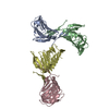

 Sample preparation
 Processing