 Movie
Movie Controller

[English] 日本語
 Yorodumi
- PDB-2nrc: C28A Mutant of Succinyl-CoA:3-Ketoacid CoA Transferase from Pig Heart -
+ Open data

- Basic information
| Entry | Database: PDB / ID: 2nrc | ||||||
|---|---|---|---|---|---|---|---|
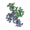
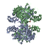
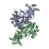
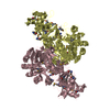
| Title | C28A Mutant of Succinyl-CoA:3-Ketoacid CoA Transferase from Pig Heart | ||||||
 Components Components | Succinyl-CoA:3-ketoacid-coenzyme A transferase 1 | ||||||
 Keywords Keywords |  TRANSFERASE / ALPHA/BETA PROTEIN TRANSFERASE / ALPHA/BETA PROTEIN | ||||||
| Function / homology |  Function and homology information Function and homology informationUtilization of Ketone Bodies / cellular ketone body metabolic process / 3-oxoacid CoA-transferase / succinyl-CoA:3-oxo-acid CoA-transferase activity / ketone body catabolic process / Mitochondrial protein degradation / protein homodimerization activity / mitochondrionSimilarity search - Function | ||||||
| Biological species |  Sus scrofa (pig) Sus scrofa (pig) | ||||||
| Method | X-RAY DIFFRACTION / SYNCHROTRON / FOURIER SYNTHESIS / Resolution: 2.05 Å | ||||||
 Authors Authors | Tammam, S.D. / Fraser, M.E. | ||||||
 Citation Citation | Journal: Biochemistry / Year: 2007 Title: Identification of the Cysteine Residue Exposed by the Conformational Change in Pig Heart Succinyl-CoA:3-Ketoacid Coenzyme A Transferase on Binding Coenzyme A. Authors: Tammam, S.D. / Rochet, J.C. / Fraser, M.E. | ||||||
| History |
|
- Structure visualization
| Structure viewer | Molecule: MolmilJmol/JSmol |
|---|
- Downloads & links
-Download
| PDBx/mmCIF format | 2nrc.cif.gz | 355.3 KB | Display | PDBx/mmCIF format |
|---|---|---|---|---|
| PDB format | pdb2nrc.ent.gz | 290.6 KB | Display | PDB format |
| PDBx/mmJSON format | 2nrc.json.gz | Tree view | PDBx/mmJSON format | |
| Others |  Other downloads Other downloads |
-Validation report
| Arichive directory | https://data.pdbj.org/pub/pdb/validation_reports/nr/2nrcftp://data.pdbj.org/pub/pdb/validation_reports/nr/2nrc | HTTPS FTP |
|---|
-Related structure data


-Links
 PDBj
PDBj- Assembly
| Deposited unit | 
| ||||||||
|---|---|---|---|---|---|---|---|---|---|
| 1 | 
| ||||||||
| 2 | 
| ||||||||
| 3 |
| ||||||||
| Unit cell |
|
-Components
| #1: Protein | Mass: 52261.895 Da / Num. of mol.: 4 / Mutation: C28A Source method: isolated from a genetically manipulated source Source: (gene. exp.) Sus scrofa (pig) / Gene: OXCT1, OXCT, SCOT / Organ: Heart / Plasmid: pT7-7 / Species (production host): Escherichia coli / Production host:  Escherichia coli BL21(DE3) (bacteria) / Strain (production host): BL21(DE3) / References: UniProt: Q29551, 3-oxoacid CoA-transferase Escherichia coli BL21(DE3) (bacteria) / Strain (production host): BL21(DE3) / References: UniProt: Q29551, 3-oxoacid CoA-transferase#2: Water | ChemComp-HOH / | Water Mass: 18.015 Da / Num. of mol.: 284 / Source method: isolated from a natural source / Formula: H2O Mass: 18.015 Da / Num. of mol.: 284 / Source method: isolated from a natural source / Formula: H2O |
|---|
-Experimental details
-Experiment
| Experiment | Method: X-RAY DIFFRACTION / Number of used crystals: 1 |
|---|
- Sample preparation
| Crystal | Density Matthews: 2.09 Å3/Da / Density % sol: 41.23 % |
|---|---|
| Crystal grow | Temperature: 287 K / Method: vapor diffusion, hanging drop / pH: 8 Details: PEG 2000, Tris-HCl, glycerol, pH 8.0, VAPOR DIFFUSION, HANGING DROP, temperature 287K |
-Data collection
| Diffraction | Mean temperature: 100 K |
|---|---|
| Diffraction source | Source: SYNCHROTRON / Site: ALS  / Beamline: 8.3.1 / Wavelength: 1.07 / Beamline: 8.3.1 / Wavelength: 1.07 |
| Detector | Type: ADSC QUANTUM 315 / Detector: CCD / Date: Jul 23, 2003 |
| Radiation | Monochromator: DOUBLE CRYSTAL / Protocol: SINGLE WAVELENGTH / Monochromatic (M) / Laue (L): M / Scattering type: x-ray |
| Radiation wavelength | Wavelength: 1.07 Å / Relative weight: 1 |
| Reflection | Resolution: 2.05→47.9 Å / Num. all: 95166 / Num. obs: 95166 / % possible obs: 92.5 % / Observed criterion σ(F): 0 / Observed criterion σ(I): 0 / Redundancy: 3.1 % / Biso Wilson estimate: 27.6 Å2 / Rmerge(I) obs: 0.113 / Net I/σ(I): 11.3 |
| Reflection shell | Resolution: 2.05→2.09 Å / Redundancy: 2.2 % / Rmerge(I) obs: 0.257 / Mean I/σ(I) obs: 1.9 / Num. unique all: 3128 / % possible all: 60.8 |
- Processing
| Software |
| |||||||||||||||||||||||||
|---|---|---|---|---|---|---|---|---|---|---|---|---|---|---|---|---|---|---|---|---|---|---|---|---|---|---|
| Refinement | Method to determine structure: FOURIER SYNTHESIS Starting model: C28S mutant Resolution: 2.05→47.9 Å / Isotropic thermal model: Isotropic / Cross valid method: THROUGHOUT / σ(F): 0 / σ(I): 0 / Stereochemistry target values: Engh & Huber
| |||||||||||||||||||||||||
| Displacement parameters |
| |||||||||||||||||||||||||
| Refine analyze |
| |||||||||||||||||||||||||
| Refinement step | Cycle: LAST / Resolution: 2.05→47.9 Å
| |||||||||||||||||||||||||
| Refine LS restraints |
| |||||||||||||||||||||||||
| LS refinement shell | Resolution: 2.06→2.15 Å / Rfactor Rfree error: 0.017
|
