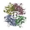
 Movie
Movie Controller


+ Open data

- Basic information
| Entry | Database: PDB / ID: 1ooy | ||||||
|---|---|---|---|---|---|---|---|
| Title | SUCCINYL-COA:3-KETOACID COA TRANSFERASE FROM PIG HEART | ||||||
 Components Components | Succinyl-CoA:3-ketoacid-coenzyme A transferase, mitochondrial precursor | ||||||
 Keywords Keywords |  TRANSFERASE / ALPHA/BETA PROTEIN TRANSFERASE / ALPHA/BETA PROTEIN | ||||||
| Function / homology |  Function and homology information Function and homology informationUtilization of Ketone Bodies / cellular ketone body metabolic process / 3-oxoacid CoA-transferase / succinyl-CoA:3-oxo-acid CoA-transferase activity / ketone body catabolic process / Mitochondrial protein degradation / protein homodimerization activity / mitochondrionSimilarity search - Function | ||||||
| Biological species |  Sus scrofa (pig) Sus scrofa (pig) | ||||||
| Method | X-RAY DIFFRACTION / SYNCHROTRON / MOLECULAR REPLACEMENT / Resolution: 1.7 Å | ||||||
 Authors Authors | Coros, A.M. / Swenson, L. / Wolodko, W.T. / Fraser, M.E. | ||||||
 Citation Citation | Journal: Acta Crystallogr.,Sect.D / Year: 2004 Title: Structure of the CoA transferase from pig heart to 1.7 A resolution. Authors: Coros, A.M. / Swenson, L. / Wolodko, W.T. / Fraser, M.E. | ||||||
| History |
|
- Structure visualization
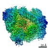
| Structure viewer | Molecule: MolmilJmol/JSmol |
|---|
- Downloads & links
-Download
| PDBx/mmCIF format | 1ooy.cif.gz | 214.2 KB | Display | PDBx/mmCIF format |
|---|---|---|---|---|
| PDB format | pdb1ooy.ent.gz | 169.5 KB | Display | PDB format |
| PDBx/mmJSON format | 1ooy.json.gz | Tree view | PDBx/mmJSON format | |
| Others |  Other downloads Other downloads |
-Validation report
| Arichive directory | https://data.pdbj.org/pub/pdb/validation_reports/oo/1ooyftp://data.pdbj.org/pub/pdb/validation_reports/oo/1ooy | HTTPS FTP |
|---|
-Related structure data
| Related structure data | 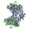 1oozC  1opeC 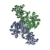 1m3eS S: Starting model for refinement C: citing same article ( |
|---|---|
| Similar structure data |


-Links
 PDBj
PDBj- Assembly
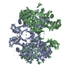
| Deposited unit | 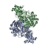
| ||||||||
|---|---|---|---|---|---|---|---|---|---|
| 1 | 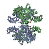
| ||||||||
| 2 |
| ||||||||
| Unit cell |
| ||||||||
| Components on special symmetry positions |
| ||||||||
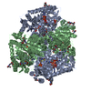
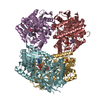
| Details | The biological assembly is a tetramer, generated by the two-fold axis: -x, -y,z. |
-Components
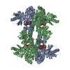
| #1: Protein | Mass: 52293.957 Da / Num. of mol.: 2 / Fragment: succinyl-CoA:3-ketoacid CoA transferase monomer Source method: isolated from a genetically manipulated source Source: (gene. exp.) Sus scrofa (pig) / Gene: OXCT OR SCOT / Plasmid: PT7-7 / Species (production host): Escherichia coli / Production host:  Escherichia coli BL21(DE3) (bacteria) / Strain (production host): BL21(DE3) / References: UniProt: Q29551, 3-oxoacid CoA-transferase Escherichia coli BL21(DE3) (bacteria) / Strain (production host): BL21(DE3) / References: UniProt: Q29551, 3-oxoacid CoA-transferase#2: Chemical | ChemComp-K /   Mass: 39.098 Da / Num. of mol.: 4 / Source method: obtained synthetically / Formula: K Mass: 39.098 Da / Num. of mol.: 4 / Source method: obtained synthetically / Formula: K#3: Chemical | ChemComp-PO4 / | Phosphate  Mass: 94.971 Da / Num. of mol.: 1 / Source method: obtained synthetically / Formula: PO4 Mass: 94.971 Da / Num. of mol.: 1 / Source method: obtained synthetically / Formula: PO4#4: Water | ChemComp-HOH / | Water Mass: 18.015 Da / Num. of mol.: 975 / Source method: isolated from a natural source / Formula: H2O Mass: 18.015 Da / Num. of mol.: 975 / Source method: isolated from a natural source / Formula: H2O |
|---|
-Experimental details
-Experiment
| Experiment | Method: X-RAY DIFFRACTION / Number of used crystals: 1 |
|---|
- Sample preparation
| Crystal | Density Matthews: 2.06 Å3/Da / Density % sol: 39.72 % |
|---|---|
| Crystal grow | Temperature: 298 K / Method: vapor diffusion, hanging drop / pH: 7.5 Details: PEG 2000, sodium/potassium phosphate, pH 7.5, VAPOR DIFFUSION, HANGING DROP, temperature 298K |
-Data collection
| Diffraction | Mean temperature: 100 K |
|---|---|
| Diffraction source | Source: SYNCHROTRON / Site: NSLS  / Beamline: X12C / Wavelength: 1.1 Å / Beamline: X12C / Wavelength: 1.1 Å |
| Detector | Type: BRANDEIS / Detector: CCD / Date: Jul 10, 1998 |
| Radiation | Monochromator: Si 111 CHANNEL / Protocol: SINGLE WAVELENGTH / Monochromatic (M) / Laue (L): M / Scattering type: x-ray |
| Radiation wavelength | Wavelength: 1.1 Å / Relative weight: 1 |
| Reflection | Resolution: 1.7→20 Å / Num. all: 111125 / Num. obs: 100807 / % possible obs: 90.7 % / Observed criterion σ(F): 0 / Observed criterion σ(I): -3.7 / Biso Wilson estimate: 15.1 Å2 / Rmerge(I) obs: 0.059 / Net I/σ(I): 20.6 |
| Reflection shell | Resolution: 1.7→1.73 Å / Rmerge(I) obs: 0.216 / Mean I/σ(I) obs: 2.5 / % possible all: 73.8 |
- Processing
| Software |
| ||||||||||||||||||||||||||||||||||||||||||||||||||||||||||||||||||||||||||||||||
|---|---|---|---|---|---|---|---|---|---|---|---|---|---|---|---|---|---|---|---|---|---|---|---|---|---|---|---|---|---|---|---|---|---|---|---|---|---|---|---|---|---|---|---|---|---|---|---|---|---|---|---|---|---|---|---|---|---|---|---|---|---|---|---|---|---|---|---|---|---|---|---|---|---|---|---|---|---|---|---|---|---|
| Refinement | Method to determine structure: MOLECULAR REPLACEMENT Starting model: PDB ENTRY 1M3E Resolution: 1.7→19.81 Å / Rfactor Rfree error: 0.002 / Isotropic thermal model: RESTRAINED / Cross valid method: THROUGHOUT / σ(F): 0 / Stereochemistry target values: Engh & Huber
| ||||||||||||||||||||||||||||||||||||||||||||||||||||||||||||||||||||||||||||||||
| Solvent computation | Solvent model: FLAT MODEL / Bsol: 39.4975 Å2 / ksol: 0.359214 e/Å3 | ||||||||||||||||||||||||||||||||||||||||||||||||||||||||||||||||||||||||||||||||
| Displacement parameters | Biso mean: 19.7 Å2
| ||||||||||||||||||||||||||||||||||||||||||||||||||||||||||||||||||||||||||||||||
| Refine analyze |
| ||||||||||||||||||||||||||||||||||||||||||||||||||||||||||||||||||||||||||||||||
| Refinement step | Cycle: LAST / Resolution: 1.7→19.81 Å
| ||||||||||||||||||||||||||||||||||||||||||||||||||||||||||||||||||||||||||||||||
| Refine LS restraints |
| ||||||||||||||||||||||||||||||||||||||||||||||||||||||||||||||||||||||||||||||||
| LS refinement shell | Resolution: 1.7→1.81 Å / Rfactor Rfree error: 0.006 / Total num. of bins used: 6
| ||||||||||||||||||||||||||||||||||||||||||||||||||||||||||||||||||||||||||||||||
| Xplor file |
|
