 Movie
Movie Controller

[English] 日本語
 Yorodumi
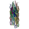
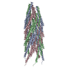
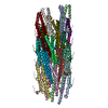
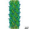
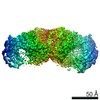
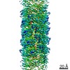
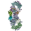
- PDB-2mjz: Capsid model of M13 bacteriophage virus from Magic-angle spinning... -
+ Open data

- Basic information
| Entry | Database: PDB / ID: 2mjz | ||||||
|---|---|---|---|---|---|---|---|
| Title | Capsid model of M13 bacteriophage virus from Magic-angle spinning NMR and Rosetta modeling | ||||||
 Components Components | Capsid protein G8P | ||||||
 Keywords Keywords | VIRAL PROTEIN / molecular assembly | ||||||
| Function / homology |  Function and homology information Function and homology information | ||||||
| Biological species |  Enterobacteria phage M13 (virus) Enterobacteria phage M13 (virus) | ||||||
| Method | SOLID-STATE NMR / Fold-and-dock | ||||||
| Model details | lowest energy, model1 | ||||||
 Authors Authors | Morag, O. / Sgourakis, N.G. / Baker, D. / Goldbourt, A. | ||||||
 Citation Citation | Journal: Proc.Natl.Acad.Sci.USA / Year: 2015 Title: The NMR-Rosetta capsid model of M13 bacteriophage reveals a quadrupled hydrophobic packing epitope. Authors: Morag, O. / Sgourakis, N.G. / Baker, D. / Goldbourt, A. | ||||||
| History |
|
- Structure visualization
| Structure viewer | Molecule: MolmilJmol/JSmol |
|---|
- Downloads & links
-Download
| PDBx/mmCIF format | 2mjz.cif.gz | 1.5 MB | Display | PDBx/mmCIF format |
|---|---|---|---|---|
| PDB format | pdb2mjz.ent.gz | 1.3 MB | Display | PDB format |
| PDBx/mmJSON format | 2mjz.json.gz | Tree view | PDBx/mmJSON format | |
| Others |  Other downloads Other downloads |
-Validation report
| Arichive directory | https://data.pdbj.org/pub/pdb/validation_reports/mj/2mjzftp://data.pdbj.org/pub/pdb/validation_reports/mj/2mjz | HTTPS FTP |
|---|
-Related structure data
| Similar structure data | |
|---|---|
| Other databases |

-Links
 PDBj
PDBj- Assembly
| Deposited unit | 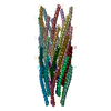
| |||||||||
|---|---|---|---|---|---|---|---|---|---|---|
| 1 |
| |||||||||
| NMR ensembles |
| |||||||||
| Details | THE ASSEMBLY REPRESENTED IN THIS ENTRY HAS A CLASS 1 SYMMETRY (C5S2). THERE IS A 5-FOLD CIRCULAR SYMMETRY AROUND THE VIRAL AXIS (Z COORDINATE) WITH THE FOLLOWING PARAMETERS: MODEL 1: ROTATION PER PENTAMER (TWIST) = 36.6 DEGREES RISE PER PENTAMER (HEIGHT) = 16.7 ANGSTROMS. MODEL 2: ROTATION PER PENTAMER (TWIST) = 36.1 DEGREES RISE PER PENTAMER (HEIGHT) = 16.7 ANGSTROMS. MODEL 3: ROTATION PER PENTAMER (TWIST) = 36.4 DEGREES RISE PER PENTAMER (HEIGHT) = 16.6 ANGSTROMS. |
-Components
| #1: Protein/peptide | / Coat protein B / Gene 8 protein / G8P / M13 procoat / Major coat protein Mass: 5243.014 Da / Num. of mol.: 35 Source method: isolated from a genetically manipulated source Source: (gene. exp.) Enterobacteria phage M13 (virus) / Strain: M13KO7 / Gene: VIII / References: UniProt: P69541 |
|---|
-Experimental details
-Experiment
| Experiment | Method: SOLID-STATE NMR Details: Solid-state NMR structure of an intact M13 bacteriophage capsid | ||||||||||||||||||||||||||||||||||||||||||||
|---|---|---|---|---|---|---|---|---|---|---|---|---|---|---|---|---|---|---|---|---|---|---|---|---|---|---|---|---|---|---|---|---|---|---|---|---|---|---|---|---|---|---|---|---|---|
| NMR experiment |
| ||||||||||||||||||||||||||||||||||||||||||||
| NMR details | Text: IN THE PDB FILE THE 35 SUBUNITS MODELED ARE REPRESENTED AS CHAINS NUMBERED FROM A-Z, a-i. THE NOTATION WE USED IN THE PAPER FOR DESCRIBING THE CAPSID ARRANGEMENT IS BASED ON THE PENTAMER ...Text: IN THE PDB FILE THE 35 SUBUNITS MODELED ARE REPRESENTED AS CHAINS NUMBERED FROM A-Z, a-i. THE NOTATION WE USED IN THE PAPER FOR DESCRIBING THE CAPSID ARRANGEMENT IS BASED ON THE PENTAMER SYMMETRY; EACH SUBUNIT PNM IS GIVEN TWO INDICES, WHERE THE INDEX N INDICATES THE PENTAMER NUMBER (N BETWEEN 1-7 WHERE N=1 CORRESPONDS TO THE C-TERMINAL PART) AND M INDICATES THE IDENTITY OF THE SUBUNIT WITHIN EACH PENTAMER(M=1-5). THE TRANSFORMATION FROM THE PAPER'S NOTATION TO THE RESPECTIVE PDB CHAINS IS AS FOLLOWS: P11 J P12 K P13 L P14 M P15 N P21 O P22 P P23 Q P24 R P25 S P31 T P32 U P33 V P34 W P35 X P41 Y P42 Z P43 a P44 b P45 c P51 d P52 e P53 f P54 g P55 h P61 i P62 A P63 B P64 C P65 D P71 E P72 F P73 G P74 H P75 I |
- Sample preparation
| Details |
| ||||||||||||||||
|---|---|---|---|---|---|---|---|---|---|---|---|---|---|---|---|---|---|
| Sample |
| ||||||||||||||||
| Sample conditions | Ionic strength: 5 / pH: 8 / Pressure: ambient / Temperature: 288 K |
-NMR measurement
| NMR spectrometer | Type: Bruker Avance III / Manufacturer: Bruker / Model: Avance III / Field strength: 600 MHz |
|---|
- Processing
| NMR software |
| ||||||||||||||||||||||||||||||||
|---|---|---|---|---|---|---|---|---|---|---|---|---|---|---|---|---|---|---|---|---|---|---|---|---|---|---|---|---|---|---|---|---|---|
| Refinement | Method: Fold-and-dock / Software ordinal: 1 Details: magic angle spinning, backbone fragment-based Monte Carlo trials followed by combinatorial sidechain packing | ||||||||||||||||||||||||||||||||
| NMR representative | Selection criteria: lowest energy | ||||||||||||||||||||||||||||||||
| NMR ensemble | Conformer selection criteria: target function / Conformers calculated total number: 5000 / Conformers submitted total number: 3 |
