 Movie
Movie Controller

[English] 日本語
 Yorodumi
- EMDB-10721: 2.85 A cryo-EM structure of the in vivo assembled type 1 pilus rod -
+ Open data

- Basic information
| Entry | Database: EMDB / ID: EMD-10721 | |||||||||
|---|---|---|---|---|---|---|---|---|---|---|
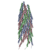
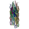
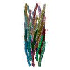
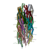
| Title | 2.85 A cryo-EM structure of the in vivo assembled type 1 pilus rod | |||||||||
 Map data Map data | ||||||||||
 Sample Sample |
| |||||||||
 Keywords Keywords | FimA /  pilus / monomer / subunit / pili / main structural subunit / high resolution / STRUCTURAL PROTEIN / cryo-EM / helical processing / RELION / Chaperone-usher pilus pilus / monomer / subunit / pili / main structural subunit / high resolution / STRUCTURAL PROTEIN / cryo-EM / helical processing / RELION / Chaperone-usher pilus | |||||||||
| Function / homology | cell adhesion involved in single-species biofilm formation / Fimbrial-type adhesion domain / Fimbrial protein / Fimbrial-type adhesion domain superfamily / Adhesion domain superfamily / pilus / cell adhesion / identical protein binding / Type-1 fimbrial protein, A chain Function and homology information Function and homology information | |||||||||
| Biological species |  Escherichia coli (E. coli) Escherichia coli (E. coli) | |||||||||
| Method | helical reconstruction / cryo EM / Resolution: 2.85 Å | |||||||||
 Authors Authors | Zyla D / Hospenthal M | |||||||||
| Funding support |  Switzerland, 2 items Switzerland, 2 items
| |||||||||
 Citation Citation | Journal: Nat Commun / Year: 2024 Title: The assembly platform FimD is required to obtain the most stable quaternary structure of type 1 pili. Authors: Dawid S Zyla / Thomas Wiegand / Paul Bachmann / Rafal Zdanowicz / Christoph Giese / Beat H Meier / Gabriel Waksman / Manuela K Hospenthal / Rudi Glockshuber /    Abstract: Type 1 pili are important virulence factors of uropathogenic Escherichia coli that mediate bacterial attachment to epithelial cells in the urinary tract. The pilus rod is comprised of thousands of ...Type 1 pili are important virulence factors of uropathogenic Escherichia coli that mediate bacterial attachment to epithelial cells in the urinary tract. The pilus rod is comprised of thousands of copies of the main structural subunit FimA and is assembled in vivo by the assembly platform FimD. Although type 1 pilus rods can self-assemble from FimA in vitro, this reaction is slower and produces structures with lower kinetic stability against denaturants compared to in vivo-assembled rods. Our study reveals that FimD-catalysed in vitro-assembled type 1 pilus rods attain a similar stability as pilus rods assembled in vivo. Employing structural, biophysical and biochemical analyses, we show that in vitro assembly reactions lacking FimD produce pilus rods with structural defects, reducing their stability against dissociation. Overall, our results indicate that FimD is not only required for the catalysis of pilus assembly, but also to control the assembly of the most stable quaternary structure. | |||||||||
| History |
|
- Structure visualization
| Movie |
Movie viewer |
|---|---|
| Structure viewer | EM map: SurfViewMolmilJmol/JSmol |
| Supplemental images |

- Downloads & links
-EMDB archive
| Map data | emd_10721.map.gz | 49.6 MB | EMDB map data format | |
|---|---|---|---|---|
| Header (meta data) | emd-10721-v30.xmlemd-10721.xml | 19.6 KB 19.6 KB | Display Display | EMDB header |
| FSC (resolution estimation) | emd_10721_fsc.xml | 9.1 KB | Display | FSC data file |
| Images | 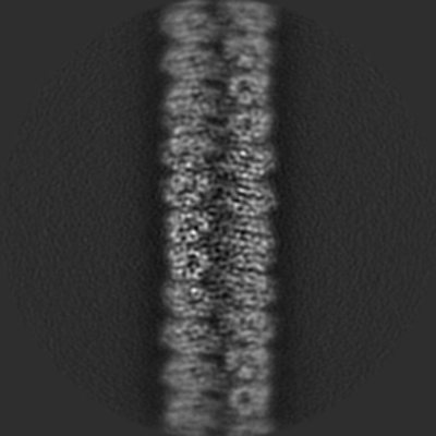 emd_10721.png emd_10721.png | 80.8 KB | ||
| Masks | emd_10721_msk_1.map | 64 MB | Mask map | |
| Filedesc metadata | emd-10721.cif.gz | 6.3 KB | ||
| Others | emd_10721_additional_1.map.gzemd_10721_half_map_1.map.gzemd_10721_half_map_2.map.gz | 4.5 MB 49.6 MB 49.6 MB | ||
| Archive directory |  http://ftp.pdbj.org/pub/emdb/structures/EMD-10721ftp://ftp.pdbj.org/pub/emdb/structures/EMD-10721 http://ftp.pdbj.org/pub/emdb/structures/EMD-10721ftp://ftp.pdbj.org/pub/emdb/structures/EMD-10721 | HTTPS FTP |
-Related structure data
| Related structure data |  6y7sMC  8psvC  8ptuC M: atomic model generated by this map C: citing same article ( |
|---|---|
| Similar structure data |


-Links
| EMDB pages | EMDB (EBI/PDBe) / EMDataResource |
|---|
-Map
| File | Download / File: emd_10721.map.gz / Format: CCP4 / Size: 64 MB / Type: IMAGE STORED AS FLOATING POINT NUMBER (4 BYTES) | ||||||||||||||||||||||||||||||||||||||||||||||||||||||||||||
|---|---|---|---|---|---|---|---|---|---|---|---|---|---|---|---|---|---|---|---|---|---|---|---|---|---|---|---|---|---|---|---|---|---|---|---|---|---|---|---|---|---|---|---|---|---|---|---|---|---|---|---|---|---|---|---|---|---|---|---|---|---|
| Voxel size | X=Y=Z: 1.08187 Å | ||||||||||||||||||||||||||||||||||||||||||||||||||||||||||||
| Density |
| ||||||||||||||||||||||||||||||||||||||||||||||||||||||||||||
| Symmetry | Space group: 1 | ||||||||||||||||||||||||||||||||||||||||||||||||||||||||||||
| Details | EMDB XML:
CCP4 map header:
| ||||||||||||||||||||||||||||||||||||||||||||||||||||||||||||
-Supplemental data
-Mask #1
| File | emd_10721_msk_1.map | ||||||||||||
|---|---|---|---|---|---|---|---|---|---|---|---|---|---|
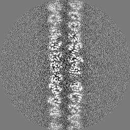
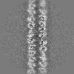
| Projections & Slices |
| ||||||||||||
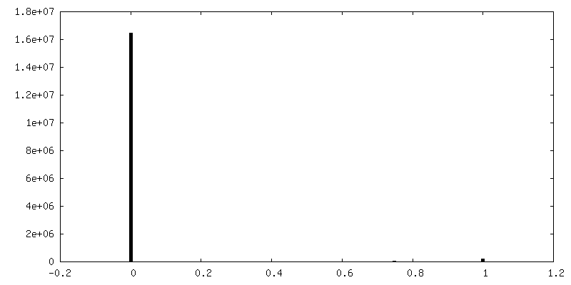
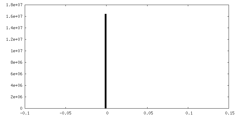
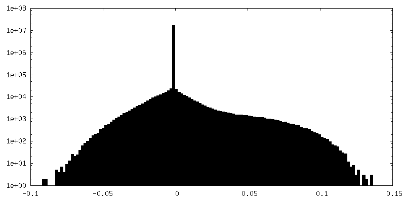
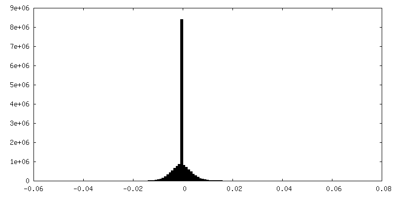
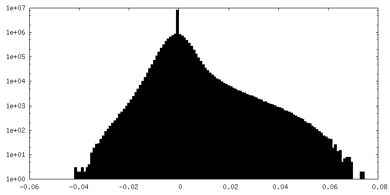
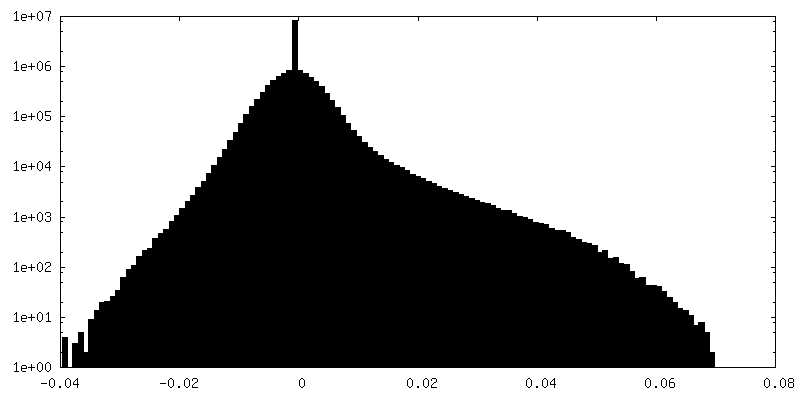
| Density Histograms |
 Z
Z Y
Y X
X


-Additional map: #1
| File | emd_10721_additional_1.map | ||||||||||||
|---|---|---|---|---|---|---|---|---|---|---|---|---|---|
| Projections & Slices |
| ||||||||||||
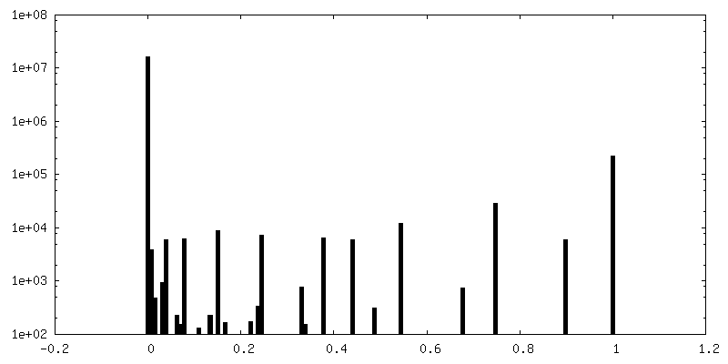
| Density Histograms |
-Half map: #2
| File | emd_10721_half_map_1.map | ||||||||||||
|---|---|---|---|---|---|---|---|---|---|---|---|---|---|
| Projections & Slices |
| ||||||||||||
| Density Histograms |
-Half map: #1
| File | emd_10721_half_map_2.map | ||||||||||||
|---|---|---|---|---|---|---|---|---|---|---|---|---|---|
| Projections & Slices |
| ||||||||||||
| Density Histograms |

- Sample components
-Entire : Type 1 pilus rod
| Entire | Name: Type 1 pilus rod |
|---|---|
| Components |
|
-Supramolecule #1: Type 1 pilus rod
| Supramolecule | Name: Type 1 pilus rod / type: complex / ID: 1 / Parent: 0 / Macromolecule list: all Details: Type 1 pili recombinantly expressed, assembled in vivo and subsequently purified from the E. coli cell surface. |
|---|---|
| Source (natural) | Organism: Escherichia coli (E. coli) |
| Molecular weight | Theoretical: 20.3 kDa/nm |
-Macromolecule #1: Type-1 fimbrial protein, A chain
| Macromolecule | Name: Type-1 fimbrial protein, A chain / type: protein_or_peptide / ID: 1 / Number of copies: 6 / Enantiomer: LEVO |
|---|---|
| Source (natural) | Organism: Escherichia coli (E. coli) |
| Molecular weight | Theoretical: 18.121074 KDa |
| Recombinant expression | Organism: Escherichia coli str. K-12 substr. W3110 (bacteria) |
| Sequence | String: MKIKTLAIVV LSALSLSSTA ALAAATTVNG GTVHFKGEVV NAACAVDAGS VDQTVQLGQV RTASLAQEGA TSSAVGFNIQ LNDCDTNVA SKAAVAFLGT AIDAGHTNVL ALQSSAAGSA TNVGVQILDR TGAALTLDGA TFSSETTLNN GTNTIPFQAR Y FATGAATP GAANADATFK VQYQ UniProtKB: Type-1 fimbrial protein, A chain |
-Experimental details
-Structure determination
| Method | cryo EM |
|---|---|
 Processing Processing | helical reconstruction |
| Aggregation state | filament |
-Sample preparation
| Concentration | 1.58 mg/mL |
|---|---|
| Buffer | pH: 7 / Details: in ddH2O. |
| Grid | Model: Quantifoil R1.2/1.3 / Material: COPPER / Mesh: 400 / Pretreatment - Type: GLOW DISCHARGE / Pretreatment - Time: 45 sec. / Pretreatment - Atmosphere: AIR |
| Vitrification | Cryogen name: ETHANE / Chamber humidity: 70 % / Chamber temperature: 277 K / Instrument: FEI VITROBOT MARK IV Details: 3 ul sample, 30 s wait time, 0.5 s drain time, 6 s blotting. |
- Electron microscopy
| Microscope | FEI TITAN KRIOS |
|---|---|
| Electron beam | Acceleration voltage: 300 kV / Electron source: FIELD EMISSION GUN |
| Electron optics | C2 aperture diameter: 70.0 µm / Illumination mode: FLOOD BEAM / Imaging mode: BRIGHT FIELDBright-field microscopy / Cs: 2.7 mm / Nominal defocus max: 3.5 µm / Nominal defocus min: 1.5 µm / Nominal magnification: 130000 |
| Sample stage | Specimen holder model: FEI TITAN KRIOS AUTOGRID HOLDER / Cooling holder cryogen: NITROGEN |
| Image recording | Film or detector model: GATAN K2 SUMMIT (4k x 4k) / Detector mode: COUNTING / Number grids imaged: 1 / Number real images: 3469 / Average exposure time: 8.0 sec. / Average electron dose: 50.4 e/Å2 |
| Experimental equipment |  Model: Titan Krios / Image courtesy: FEI Company |
-Image processing
| Segment selection | Number selected: 516000 Details: Autopicking based on the 2D classes from manually chosen filaments |
|---|---|
| Startup model | Type of model: OTHER / Details: featureless cylinder |
| Final angle assignment | Type: NOT APPLICABLE / Software - Name: RELION (ver. 3.08) |
| Final reconstruction | Number classes used: 1 Applied symmetry - Helical parameters - Δz: 7.85334 Å Applied symmetry - Helical parameters - Δ&Phi: 115.001 ° Applied symmetry - Helical parameters - Axial symmetry: C1 (asymmetric) Algorithm: FOURIER SPACE / Resolution.type: BY AUTHOR / Resolution: 2.85 Å / Resolution method: FSC 0.143 CUT-OFF / Software - Name: RELION (ver. 3.08) / Details: local searches 0.9 degree with mask / Number images used: 40805 |
| FSC plot (resolution estimation) | 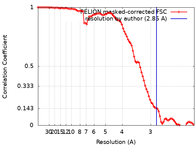 |
-Atomic model buiding 1
| Initial model | PDB ID: Chain - Chain ID: D / Chain - Source name: PDB / Chain - Initial model type: experimental model |
|---|---|
| Refinement | Space: REAL / Protocol: RIGID BODY FIT / Overall B value: 49.9714 / Target criteria: Correlation coefficient |
| Output model | PDB-6y7s: |

