 Movie
Movie Controller


+ Open data

- Basic information
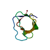
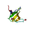
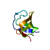
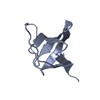
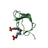
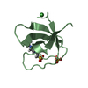
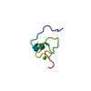
| Entry | Database: PDB / ID: 2lp5 | ||||||
|---|---|---|---|---|---|---|---|
| Title | Native Structure of the Fyn SH3 A39V/N53P/V55L | ||||||
 Components Components | Tyrosine-protein kinase Fyn | ||||||
 Keywords Keywords |  TRANSFERASE / amyloid fibril / protein folding intermediate TRANSFERASE / amyloid fibril / protein folding intermediate | ||||||
| Function / homology |  Function and homology information Function and homology informationforebrain development / extrinsic component of cytoplasmic side of plasma membrane / tubulin binding / cell surface receptor protein tyrosine kinase signaling pathway / non-specific protein-tyrosine kinase / neuron migration / non-membrane spanning protein tyrosine kinase activity / peptidyl-tyrosine phosphorylation / regulation of cell shape / perikaryon ...forebrain development / extrinsic component of cytoplasmic side of plasma membrane / tubulin binding / cell surface receptor protein tyrosine kinase signaling pathway / non-specific protein-tyrosine kinase / neuron migration / non-membrane spanning protein tyrosine kinase activity / peptidyl-tyrosine phosphorylation / regulation of cell shape / perikaryon / protein tyrosine kinase activity / cell differentiation / protein kinase activity / membrane raft / signaling receptor binding / innate immune response / ATP binding / metal ion binding / nucleus / cytosolSimilarity search - Function | ||||||
| Biological species |  Gallus gallus (chicken) Gallus gallus (chicken) | ||||||
| Method | SOLUTION NMR / simulated annealing | ||||||
| Model details | lowest energy, model 1 | ||||||
 Authors Authors | Neudecker, P. / Robustelli, P. / Cavalli, A. / Vendruscolo, M. / Kay, L.E. | ||||||
 Citation Citation | Journal: Science / Year: 2012 Title: Structure of an intermediate state in protein folding and aggregation. Authors: Neudecker, P. / Robustelli, P. / Cavalli, A. / Walsh, P. / Lundstrom, P. / Zarrine-Afsar, A. / Sharpe, S. / Vendruscolo, M. / Kay, L.E. | ||||||
| History |
| ||||||
| Remark 650 | HELIX DETERMINATION METHOD: AUTHOR DETERMINED | ||||||
| Remark 700 | SHEET DETERMINATION METHOD: AUTHOR DETERMINED |
- Structure visualization
| Structure viewer | Molecule: MolmilJmol/JSmol |
|---|
- Downloads & links
-Download
| PDBx/mmCIF format | 2lp5.cif.gz | 183.2 KB | Display | PDBx/mmCIF format |
|---|---|---|---|---|
| PDB format | pdb2lp5.ent.gz | 151.1 KB | Display | PDB format |
| PDBx/mmJSON format | 2lp5.json.gz | Tree view | PDBx/mmJSON format | |
| Others |  Other downloads Other downloads |
-Validation report
| Arichive directory | https://data.pdbj.org/pub/pdb/validation_reports/lp/2lp5ftp://data.pdbj.org/pub/pdb/validation_reports/lp/2lp5 | HTTPS FTP |
|---|
-Related structure data
| Related structure data |  2l2pC C: citing same article ( |
|---|---|
| Similar structure data | |
| Other databases |
|


-Links
 PDBj
PDBj
- Assembly
| Deposited unit | 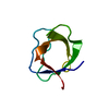
| |||||||||
|---|---|---|---|---|---|---|---|---|---|---|
| 1 |
| |||||||||
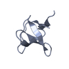
| NMR ensembles |
|
-Components
| #1: Protein | Mass: 7538.242 Da / Num. of mol.: 1 / Fragment: SH3 domain residues 85-142 / Mutation: A39V, N53P, V55L Source method: isolated from a genetically manipulated source Source: (gene. exp.) Gallus gallus (chicken) / Gene: FYN / Production host:  Escherichia coli (E. coli) / Strain (production host): BL21(DE3) Escherichia coli (E. coli) / Strain (production host): BL21(DE3)References: UniProt: Q05876, non-specific protein-tyrosine kinase |
|---|
-Experimental details
-Experiment
| Experiment | Method: SOLUTION NMR Details: Solution structure of the native Fyn SH3 domain mutant A39V/N53P/V55L from G. gallus | ||||||||||||||||||||||||||||||||||||||||||||||||||||||||||||||||||||||||||||||||||||||||||||||||||||||||||||||||||||||||||||||||||||||||||||||||||||||||||||
|---|---|---|---|---|---|---|---|---|---|---|---|---|---|---|---|---|---|---|---|---|---|---|---|---|---|---|---|---|---|---|---|---|---|---|---|---|---|---|---|---|---|---|---|---|---|---|---|---|---|---|---|---|---|---|---|---|---|---|---|---|---|---|---|---|---|---|---|---|---|---|---|---|---|---|---|---|---|---|---|---|---|---|---|---|---|---|---|---|---|---|---|---|---|---|---|---|---|---|---|---|---|---|---|---|---|---|---|---|---|---|---|---|---|---|---|---|---|---|---|---|---|---|---|---|---|---|---|---|---|---|---|---|---|---|---|---|---|---|---|---|---|---|---|---|---|---|---|---|---|---|---|---|---|---|---|---|---|
| NMR experiment |
| ||||||||||||||||||||||||||||||||||||||||||||||||||||||||||||||||||||||||||||||||||||||||||||||||||||||||||||||||||||||||||||||||||||||||||||||||||||||||||||
| NMR details | Text: The CPMG NMR relaxation dispersion experiments were supplemented with sign determination experiments as appropriate. |
- Sample preparation
| Details |
| |||||||||||||||||||||||||||||||||||||||||||||||||||||||||||||||||||||||||||||||||||||||||||||||||||||||||||||||||||||||||||||||||||||||||||||||||||||||||||||||||||||||||||||||||||||||||||||||||||||||||
|---|---|---|---|---|---|---|---|---|---|---|---|---|---|---|---|---|---|---|---|---|---|---|---|---|---|---|---|---|---|---|---|---|---|---|---|---|---|---|---|---|---|---|---|---|---|---|---|---|---|---|---|---|---|---|---|---|---|---|---|---|---|---|---|---|---|---|---|---|---|---|---|---|---|---|---|---|---|---|---|---|---|---|---|---|---|---|---|---|---|---|---|---|---|---|---|---|---|---|---|---|---|---|---|---|---|---|---|---|---|---|---|---|---|---|---|---|---|---|---|---|---|---|---|---|---|---|---|---|---|---|---|---|---|---|---|---|---|---|---|---|---|---|---|---|---|---|---|---|---|---|---|---|---|---|---|---|---|---|---|---|---|---|---|---|---|---|---|---|---|---|---|---|---|---|---|---|---|---|---|---|---|---|---|---|---|---|---|---|---|---|---|---|---|---|---|---|---|---|---|---|---|---|
| Sample |
|