 Movie
Movie Controller


+ Open data

- Basic information
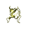
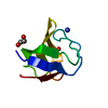
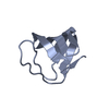
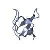
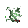
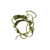
| Entry | Database: PDB / ID: 3cqt | ||||||
|---|---|---|---|---|---|---|---|
| Title | N53I V55L MUTANT of FYN SH3 DOMAIN | ||||||
 Components Components | Proto-oncogene tyrosine-protein kinase Fyn | ||||||
 Keywords Keywords |  TRANSFERASE / beta barrel / ATP-binding / Developmental protein / Kinase / Lipoprotein / Manganese / Metal-binding / Myristate / Nucleotide-binding / Palmitate / Phosphoprotein / Proto-oncogene / SH2 domain / SH3 domain / Tyrosine-protein kinase TRANSFERASE / beta barrel / ATP-binding / Developmental protein / Kinase / Lipoprotein / Manganese / Metal-binding / Myristate / Nucleotide-binding / Palmitate / Phosphoprotein / Proto-oncogene / SH2 domain / SH3 domain / Tyrosine-protein kinase | ||||||
| Function / homology |  Function and homology information Function and homology informationforebrain development / extrinsic component of cytoplasmic side of plasma membrane / tubulin binding / cell surface receptor protein tyrosine kinase signaling pathway / non-specific protein-tyrosine kinase / neuron migration / non-membrane spanning protein tyrosine kinase activity / peptidyl-tyrosine phosphorylation / regulation of cell shape / perikaryon ...forebrain development / extrinsic component of cytoplasmic side of plasma membrane / tubulin binding / cell surface receptor protein tyrosine kinase signaling pathway / non-specific protein-tyrosine kinase / neuron migration / non-membrane spanning protein tyrosine kinase activity / peptidyl-tyrosine phosphorylation / regulation of cell shape / perikaryon / protein tyrosine kinase activity / cell differentiation / protein kinase activity / membrane raft / signaling receptor binding / innate immune response / ATP binding / metal ion binding / nucleus / cytosolSimilarity search - Function | ||||||
| Biological species |  Gallus gallus (chicken) Gallus gallus (chicken) | ||||||
| Method | X-RAY DIFFRACTION / MOLECULAR REPLACEMENT / Resolution: 1.6 Å | ||||||
 Authors Authors | Neculai, A.M. / Zarrine-Afsar, A. / Howell, P.L. / Davidson, A. / Chan, H.S. | ||||||
 Citation Citation | Journal: Proc.Natl.Acad.Sci.Usa / Year: 2008 Title: Theoretical and experimental demonstration of the importance of specific nonnative interactions in protein folding. Authors: Zarrine-Afsar, A. / Wallin, S. / Neculai, A.M. / Neudecker, P. / Howell, P.L. / Davidson, A.R. / Chan, H.S. | ||||||
| History |
|
- Structure visualization
| Structure viewer | Molecule: MolmilJmol/JSmol |
|---|
- Downloads & links
-Download
| PDBx/mmCIF format | 3cqt.cif.gz | 27 KB | Display | PDBx/mmCIF format |
|---|---|---|---|---|
| PDB format | pdb3cqt.ent.gz | 16.5 KB | Display | PDB format |
| PDBx/mmJSON format | 3cqt.json.gz | Tree view | PDBx/mmJSON format | |
| Others |  Other downloads Other downloads |
-Validation report
| Arichive directory | https://data.pdbj.org/pub/pdb/validation_reports/cq/3cqtftp://data.pdbj.org/pub/pdb/validation_reports/cq/3cqt | HTTPS FTP |
|---|
-Related structure data
| Related structure data |  1fynS S: Starting model for refinement |
|---|---|
| Similar structure data |


-Links
 PDBj
PDBj- Assembly
| Deposited unit | 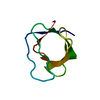
| ||||||||
|---|---|---|---|---|---|---|---|---|---|
| 1 |
| ||||||||
| Unit cell |
|
-Components
| #1: Protein | Mass: 9337.112 Da / Num. of mol.: 1 / Fragment: Fyn SH3 domain / Mutation: N53I, V55L Source method: isolated from a genetically manipulated source Source: (gene. exp.) Gallus gallus (chicken) / Production host:  Escherichia coli (E. coli) / Strain (production host): Bl21* Escherichia coli (E. coli) / Strain (production host): Bl21*References: UniProt: Q05876, non-specific protein-tyrosine kinase |
|---|---|
| #2: Water | ChemComp-HOH / Water Mass: 18.015 Da / Num. of mol.: 89 / Source method: isolated from a natural source / Formula: H2O Mass: 18.015 Da / Num. of mol.: 89 / Source method: isolated from a natural source / Formula: H2O |
-Experimental details
-Experiment
| Experiment | Method: X-RAY DIFFRACTION / Number of used crystals: 1 |
|---|
- Sample preparation
| Crystal | Density Matthews: 1.88 Å3/Da / Density % sol: 34.42 % |
|---|---|
| Crystal grow | Temperature: 298 K / Method: vapor diffusion, hanging drop / pH: 6.5 Details: 0.1 M Sodium Cacodylate, 1.4 M Sodium Acetate, pH 6.5, VAPOR DIFFUSION, HANGING DROP, temperature 298K |
-Data collection
| Diffraction | Mean temperature: 93 K |
|---|---|
| Diffraction source | Source: ROTATING ANODE / Type: RIGAKU RUH3R / Wavelength: 1.54 Å |
| Detector | Type: RIGAKU RAXIS IV / Detector: IMAGE PLATE / Date: Jul 4, 2007 / Details: mirrors |
| Radiation | Monochromator: Ni MIRROR 0.3 + Ni FILTER / Protocol: SINGLE WAVELENGTH / Monochromatic (M) / Laue (L): M / Scattering type: x-ray |
| Radiation wavelength | Wavelength: 1.54 Å / Relative weight: 1 |
| Reflection | Resolution: 1.6→45.06 Å / Num. all: 9568 / Num. obs: 8962 / % possible obs: 93.7 % / Observed criterion σ(F): 2 / Observed criterion σ(I): 2 / Redundancy: 3.4 % / Rmerge(I) obs: 0.0728 / Rsym value: 0.0175 / Net I/σ(I): 53.07 |
| Reflection shell | Resolution: 1.6→1.7 Å / Redundancy: 3.4 % / Rmerge(I) obs: 0.1178 / Mean I/σ(I) obs: 33.05 / Rsym value: 0.031 / % possible all: 97.8 |
- Processing
| Software |
| ||||||||||||||||||||||||||||||||||||||||||||||||||||||||||||||||||||||||||||||||||||||||||||||||||||||||||||||||||||||||||||||||||||||||||||||||||||||||||||||||||||||||||
|---|---|---|---|---|---|---|---|---|---|---|---|---|---|---|---|---|---|---|---|---|---|---|---|---|---|---|---|---|---|---|---|---|---|---|---|---|---|---|---|---|---|---|---|---|---|---|---|---|---|---|---|---|---|---|---|---|---|---|---|---|---|---|---|---|---|---|---|---|---|---|---|---|---|---|---|---|---|---|---|---|---|---|---|---|---|---|---|---|---|---|---|---|---|---|---|---|---|---|---|---|---|---|---|---|---|---|---|---|---|---|---|---|---|---|---|---|---|---|---|---|---|---|---|---|---|---|---|---|---|---|---|---|---|---|---|---|---|---|---|---|---|---|---|---|---|---|---|---|---|---|---|---|---|---|---|---|---|---|---|---|---|---|---|---|---|---|---|---|---|---|---|
| Refinement | Method to determine structure: MOLECULAR REPLACEMENT Starting model: PDB entry 1fyn Resolution: 1.6→45.06 Å / Cor.coef. Fo:Fc: 0.939 / Cor.coef. Fo:Fc free: 0.931 / SU B: 1.464 / SU ML: 0.055 / Cross valid method: THROUGHOUT / ESU R: 0.106 / ESU R Free: 0.103 / Stereochemistry target values: MAXIMUM LIKELIHOOD
| ||||||||||||||||||||||||||||||||||||||||||||||||||||||||||||||||||||||||||||||||||||||||||||||||||||||||||||||||||||||||||||||||||||||||||||||||||||||||||||||||||||||||||
| Solvent computation | Ion probe radii: 0.8 Å / Shrinkage radii: 0.8 Å / VDW probe radii: 1.2 Å / Solvent model: MASK | ||||||||||||||||||||||||||||||||||||||||||||||||||||||||||||||||||||||||||||||||||||||||||||||||||||||||||||||||||||||||||||||||||||||||||||||||||||||||||||||||||||||||||
| Displacement parameters | Biso mean: 15.398 Å2
| ||||||||||||||||||||||||||||||||||||||||||||||||||||||||||||||||||||||||||||||||||||||||||||||||||||||||||||||||||||||||||||||||||||||||||||||||||||||||||||||||||||||||||
| Refinement step | Cycle: LAST / Resolution: 1.6→45.06 Å
| ||||||||||||||||||||||||||||||||||||||||||||||||||||||||||||||||||||||||||||||||||||||||||||||||||||||||||||||||||||||||||||||||||||||||||||||||||||||||||||||||||||||||||
| Refine LS restraints |
| ||||||||||||||||||||||||||||||||||||||||||||||||||||||||||||||||||||||||||||||||||||||||||||||||||||||||||||||||||||||||||||||||||||||||||||||||||||||||||||||||||||||||||
| LS refinement shell | Resolution: 1.6→1.642 Å / Total num. of bins used: 20
|
