 Movie
Movie Controller

[English] 日本語
 Yorodumi
- PDB-2lm7: NMR structure of the C-terminal domain of VP7 in membrane mimicki... -
+ Open data

- Basic information
| Entry | Database: PDB / ID: 2lm7 | ||||||
|---|---|---|---|---|---|---|---|
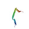
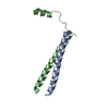
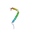
| Title | NMR structure of the C-terminal domain of VP7 in membrane mimicking micelles | ||||||
 Components Components | Outer capsid glycoprotein VP7 | ||||||
 Keywords Keywords | VIRAL PROTEIN / alpha helix / amphipathic / perforating peptide | ||||||
| Function / homology |  Function and homology information Function and homology informationhost cell endoplasmic reticulum lumen / T=13 icosahedral viral capsid / viral outer capsid / viral process / metal ion binding Similarity search - Function | ||||||
| Biological species |  Rotavirus A Rotavirus A | ||||||
| Method | SOLUTION NMR / DGSA-distance geometry simulated annealing | ||||||
| Model details | lowest energy, model 1 | ||||||
 Authors Authors | Elaid, S. / Libersou, S. / Ouldali, M. / Morellet, N. / Lepault, J. / Bouaziz, S. | ||||||
 Citation Citation | Journal: To be Published Title: NMR structure of the C-terminal domain of VP7 in membrane mimicking micelles Authors: Elaid, S. / Libersou, S. / Ouldali, M. / Rhayyat, R. / Henry, C. / Morellet, N. / Lepault, J. / Bouaziz, S. | ||||||
| History |
|
- Structure visualization
| Structure viewer | Molecule: MolmilJmol/JSmol |
|---|
- Downloads & links
-Download
| PDBx/mmCIF format | 2lm7.cif.gz | 167.8 KB | Display | PDBx/mmCIF format |
|---|---|---|---|---|
| PDB format | pdb2lm7.ent.gz | 137.9 KB | Display | PDB format |
| PDBx/mmJSON format | 2lm7.json.gz | Tree view | PDBx/mmJSON format | |
| Others |  Other downloads Other downloads |
-Validation report
| Summary document | 2lm7_validation.pdf.gz | 368.8 KB | Display | wwPDB validaton report |
|---|---|---|---|---|
| Full document | 2lm7_full_validation.pdf.gz | 391.5 KB | Display | |
| Data in XML | 2lm7_validation.xml.gz | 10.3 KB | Display | |
| Data in CIF | 2lm7_validation.cif.gz | 15.5 KB | Display | |
| Arichive directory | https://data.pdbj.org/pub/pdb/validation_reports/lm/2lm7ftp://data.pdbj.org/pub/pdb/validation_reports/lm/2lm7 | HTTPS FTP |
-Related structure data
| Related structure data | |
|---|---|
| Similar structure data |

-Links
 PDBj
PDBj
- Assembly
| Deposited unit | 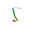
| |||||||||
|---|---|---|---|---|---|---|---|---|---|---|
| 1 |
| |||||||||
| NMR ensembles |
|
-Components
| #1: Protein | Mass: 7341.409 Da / Num. of mol.: 1 / Fragment: UNP residues 266-326 Source method: isolated from a genetically manipulated source Source: (gene. exp.) Rotavirus A / Strain: isolate Human/Belgium/4106/2000 G3-P11[14] / References: UniProt: Q3ZK60 |
|---|
-Experimental details
-Experiment
| Experiment | Method: SOLUTION NMR | ||||||||||||||||
|---|---|---|---|---|---|---|---|---|---|---|---|---|---|---|---|---|---|
| NMR experiment |
|
- Sample preparation
| Details | Contents: 1 mM VP7-61-1, 100 mM [U-100% 2H] DPC-2, 95% H2O/5% D2O Solvent system: 95% H2O/5% D2O | ||||||||||||
|---|---|---|---|---|---|---|---|---|---|---|---|---|---|
| Sample |
| ||||||||||||
| Sample conditions | Ionic strength: 0 / pH: 3 / Pressure: ambient / Temperature: 323 K |
-NMR measurement
| NMR spectrometer | Type: Bruker Avance / Manufacturer: Bruker / Model: AVANCE / Field strength: 600 MHz |
|---|
- Processing
| NMR software |
| ||||||||||||
|---|---|---|---|---|---|---|---|---|---|---|---|---|---|
| Refinement | Method: DGSA-distance geometry simulated annealing / Software ordinal: 1 | ||||||||||||
| NMR constraints | NOE constraints total: 1210 / NOE intraresidue total count: 879 / NOE long range total count: 0 / NOE medium range total count: 119 / NOE sequential total count: 212 | ||||||||||||
| NMR representative | Selection criteria: lowest energy | ||||||||||||
| NMR ensemble | Conformer selection criteria: structures with the lowest energy Conformers calculated total number: 100 / Conformers submitted total number: 8 |
