 Movie
Movie Controller

[English] 日本語
 Yorodumi
- PDB-2kwa: 1H, 13C and 15N backbone and side chain resonance assignments of ... -
+ Open data

- Basic information
| Entry | Database: PDB / ID: 2kwa | ||||||
|---|---|---|---|---|---|---|---|
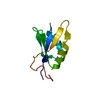
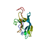
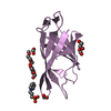
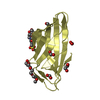
| Title | 1H, 13C and 15N backbone and side chain resonance assignments of the N-terminal domain of the histidine kinase inhibitor KipI from Bacillus subtilis | ||||||
 Components Components | Kinase A inhibitor | ||||||
 Keywords Keywords | TRANSFERASE INHIBITOR / Bacterial signal transduction / KipI / histidine kinase inhibition /  Bacillus subtilis Bacillus subtilis | ||||||
| Function / homology |  Function and homology information5-oxoprolinase (ATP-hydrolysing) / 5-oxoprolinase (ATP-hydrolyzing) activity / sporulation resulting in formation of a cellular spore / protein kinase inhibitor activity / ATP binding Function and homology information5-oxoprolinase (ATP-hydrolysing) / 5-oxoprolinase (ATP-hydrolyzing) activity / sporulation resulting in formation of a cellular spore / protein kinase inhibitor activity / ATP bindingSimilarity search - Function | ||||||
| Biological species |  Bacillus subtilis (bacteria) Bacillus subtilis (bacteria) | ||||||
| Method | SOLUTION NMR / molecular dynamics, simulated annealing | ||||||
| Model details | lowest energy, model 1 | ||||||
 Authors Authors | Hynson, R.M.G. / Kwan, A. / Jacques, D.A. / Mackay, J.P. / Trewhella, J. | ||||||
 Citation Citation | Journal: J.Mol.Biol. / Year: 2010 Title: A Novel Structure of an Antikinase and its Inhibitor Authors: Jacques, D.A. / Langley, D.B. / Hynson, R.M.G. / Whitten, A.E. / Kwan, A. / Guss, J.M. / Trewhella, J. | ||||||
| History |
|
- Structure visualization
| Structure viewer | Molecule: MolmilJmol/JSmol |
|---|
- Downloads & links
-Download
| PDBx/mmCIF format | 2kwa.cif.gz | 616.1 KB | Display | PDBx/mmCIF format |
|---|---|---|---|---|
| PDB format | pdb2kwa.ent.gz | 512.7 KB | Display | PDB format |
| PDBx/mmJSON format | 2kwa.json.gz | Tree view | PDBx/mmJSON format | |
| Others |  Other downloads Other downloads |
-Validation report
| Arichive directory | https://data.pdbj.org/pub/pdb/validation_reports/kw/2kwaftp://data.pdbj.org/pub/pdb/validation_reports/kw/2kwa | HTTPS FTP |
|---|
-Related structure data
| Similar structure data |
|---|


-Links
 PDBj
PDBj

- Assembly
| Deposited unit | 
| |||||||||
|---|---|---|---|---|---|---|---|---|---|---|
| 1 |
| |||||||||
| NMR ensembles |
|
-Components
| #1: Protein | Mass: 11565.928 Da / Num. of mol.: 1 / Fragment: N-terminal domain Source method: isolated from a genetically manipulated source Source: (gene. exp.) Bacillus subtilis (bacteria) / Strain: 168 / Gene: kipI / Production host: Escherichia coli (E. coli) / References: UniProt: P60495 |
|---|
-Experimental details
-Experiment
| Experiment | Method: SOLUTION NMR | ||||||||||||||||||||||||||||||||||||||||||||||||||||||||||||||||||||||||||||||||
|---|---|---|---|---|---|---|---|---|---|---|---|---|---|---|---|---|---|---|---|---|---|---|---|---|---|---|---|---|---|---|---|---|---|---|---|---|---|---|---|---|---|---|---|---|---|---|---|---|---|---|---|---|---|---|---|---|---|---|---|---|---|---|---|---|---|---|---|---|---|---|---|---|---|---|---|---|---|---|---|---|---|
| NMR experiment |
|
- Sample preparation
| Details | Contents: 1mM [U-100% 15N] KipI-N-1, 1mM [U-100% 13C; U-100% 15N] KipI-N-2, 1mM KipI-N-3, 90% H2O/10% D2O Solvent system: 90% H2O/10% D2O | ||||||||||||||||
|---|---|---|---|---|---|---|---|---|---|---|---|---|---|---|---|---|---|
| Sample |
| ||||||||||||||||
| Sample conditions | Ionic strength: 0.2 / pH: 6.2 / Pressure: ambient / Temperature: 298 K |
-NMR measurement
| NMR spectrometer | Type: Bruker Avance / Manufacturer: Bruker / Model: AVANCE / Field strength: 600 MHz |
|---|
- Processing
| NMR software |
| ||||||||||||||||||||||||||||||||
|---|---|---|---|---|---|---|---|---|---|---|---|---|---|---|---|---|---|---|---|---|---|---|---|---|---|---|---|---|---|---|---|---|---|
| Refinement | Method: molecular dynamics, simulated annealing / Software ordinal: 1 Details: Heating and cooling steps changed from 10000, 5000 & 4000 to 20000, 20000 & 16000 | ||||||||||||||||||||||||||||||||
| NMR constraints | Protein phi angle constraints total count: 68 / Protein psi angle constraints total count: 68 | ||||||||||||||||||||||||||||||||
| NMR representative | Selection criteria: lowest energy | ||||||||||||||||||||||||||||||||
| NMR ensemble | Conformer selection criteria: structures with the lowest energy Conformers calculated total number: 500 / Conformers submitted total number: 20 / Maximum torsion angle constraint violation: 0 ° / Representative conformer: 1 / Torsion angle constraint violation method: ARIA 2.1 |
