 ムービー
ムービー コントローラー


+ データを開く

- 基本情報
基本情報
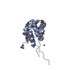
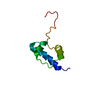
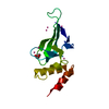
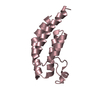
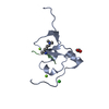
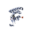
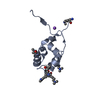
| 登録情報 | データベース: PDB / ID: 2kq6 | ||||||
|---|---|---|---|---|---|---|---|
| タイトル | The structure of the EF-hand domain of polycystin-2 suggests a mechanism for Ca2+-dependent regulation of polycystin-2 channel activity | ||||||
 要素 要素 | Polycystin-2 Polycystin 2 Polycystin 2 | ||||||
 キーワード キーワード | TRANSPORT PROTEIN (運搬体タンパク質) / Protein X / Calcium (カルシウム) / Coiled coil (コイルドコイル) / Disease mutation / Glycoprotein (糖タンパク質) / Ion transport / Ionic channel (イオンチャネル) / Membrane (生体膜) / Phosphoprotein / Polymorphism / Transmembrane (膜貫通型タンパク質) / Transport | ||||||
| 機能・相同性 |  機能・相同性情報 機能・相同性情報detection of nodal flow / metanephric smooth muscle tissue development / metanephric cortex development / metanephric cortical collecting duct development / metanephric distal tubule development / polycystin complex / mesonephric tubule development / mesonephric duct development / : / metanephric part of ureteric bud development ...detection of nodal flow / metanephric smooth muscle tissue development / metanephric cortex development / metanephric cortical collecting duct development / metanephric distal tubule development / polycystin complex / mesonephric tubule development / mesonephric duct development / : / metanephric part of ureteric bud development / determination of liver left/right asymmetry / renal tubule morphogenesis / metanephric ascending thin limb development / HLH domain binding / basal cortex / metanephric mesenchyme development / metanephric S-shaped body morphogenesis / renal artery morphogenesis / positive regulation of inositol 1,4,5-trisphosphate-sensitive calcium-release channel activity / migrasome / cilium organization / VxPx cargo-targeting to cilium / detection of mechanical stimulus / regulation of calcium ion import / cation channel complex / calcium-induced calcium release activity / muscle alpha-actinin binding / placenta blood vessel development / voltage-gated monoatomic ion channel activity / cellular response to hydrostatic pressure / voltage-gated monoatomic cation channel activity / cellular response to fluid shear stress / non-motile cilium / cellular response to osmotic stress / outward rectifier potassium channel activity / actinin binding / 繊毛 / transcription regulator inhibitor activity / inorganic cation transmembrane transport / determination of left/right symmetry / aorta development / neural tube development / voltage-gated sodium channel activity / protein heterotetramerization / ciliary membrane / branching involved in ureteric bud morphogenesis / negative regulation of G1/S transition of mitotic cell cycle / spinal cord development / heart looping / voltage-gated potassium channel activity / potassium channel activity / cytoplasmic side of endoplasmic reticulum membrane / cell surface receptor signaling pathway via JAK-STAT / centrosome duplication / negative regulation of ryanodine-sensitive calcium-release channel activity / sodium ion transmembrane transport / voltage-gated calcium channel activity / embryonic placenta development / monoatomic cation channel activity / cellular response to cAMP / release of sequestered calcium ion into cytosol / potassium ion transmembrane transport / cellular response to calcium ion / cytoskeletal protein binding / basal plasma membrane / ciliary basal body / liver development / establishment of localization in cell / lumenal side of endoplasmic reticulum membrane / calcium ion transmembrane transport / phosphoprotein binding / protein tetramerization / cytoplasmic vesicle membrane / 繊毛 / 紡錘体 / Wntシグナル経路 / cellular response to reactive oxygen species / intracellular calcium ion homeostasis / calcium ion transport / positive regulation of nitric oxide biosynthetic process / cell-cell junction / lamellipodium / heart development / regulation of cell population proliferation / ATPase binding / positive regulation of cytosolic calcium ion concentration / basolateral plasma membrane / protein homotetramerization / transmembrane transporter binding / regulation of cell cycle / negative regulation of cell population proliferation / signaling receptor binding / calcium ion binding / endoplasmic reticulum membrane / positive regulation of gene expression / ゴルジ体 / 小胞体 / protein homodimerization activity / positive regulation of transcription by RNA polymerase II / extracellular exosome類似検索 - 分子機能 | ||||||
| 生物種 |  Homo sapiens (ヒト) Homo sapiens (ヒト) | ||||||
| 手法 | 溶液NMR / XPLOR-NIH | ||||||
 データ登録者 データ登録者 | Petri, E.T. / Celic, A. / Kennedy, S.D. / Ehrlich, B.E. / Boggon, T.J. / Hodsdon, M.E. | ||||||
 引用 引用 | ジャーナル: Proc.Natl.Acad.Sci.USA / 年: 2010 タイトル: Structure of the EF-hand domain of polycystin-2 suggests a mechanism for Ca2+-dependent regulation of polycystin-2 channel activity. 著者: Petri, E.T. / Celic, A. / Kennedy, S.D. / Ehrlich, B.E. / Boggon, T.J. / Hodsdon, M.E. | ||||||
| 履歴 |
|
- 構造の表示
構造の表示
| 構造ビューア | 分子: MolmilJmol/JSmol |
|---|
- ダウンロードとリンク
-ダウンロード
| PDBx/mmCIF形式 | 2kq6.cif.gz | 468.8 KB | 表示 | PDBx/mmCIF形式 |
|---|---|---|---|---|
| PDB形式 | pdb2kq6.ent.gz | 392.3 KB | 表示 | PDB形式 |
| PDBx/mmJSON形式 | 2kq6.json.gz | ツリー表示 | PDBx/mmJSON形式 | |
| その他 |  その他のダウンロード その他のダウンロード |
-検証レポート
| アーカイブディレクトリ | https://data.pdbj.org/pub/pdb/validation_reports/kq/2kq6ftp://data.pdbj.org/pub/pdb/validation_reports/kq/2kq6 | HTTPS FTP |
|---|
-関連構造データ
| 類似構造データ | |
|---|---|
| その他のデータベース |

-リンク
 PDBj
PDBj
- 集合体
集合体
| 登録構造単位 | 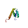
| |||||||||
|---|---|---|---|---|---|---|---|---|---|---|
| 1 |
| |||||||||
| NMR アンサンブル |
|
-要素
| #1: タンパク質 | Polycystin 2 / Polycystic kidney disease 2 protein / Autosomal dominant polycystic kidney disease type II protein ...Polycystic kidney disease 2 protein / Autosomal dominant polycystic kidney disease type II protein / Polycystwin / R48321 分子量: 9009.580 Da / 分子数: 1 / 断片: UNP residues 720-797 / 由来タイプ: 組換発現 / 由来: (組換発現) Homo sapiens (ヒト) / 遺伝子: PKD2 / 発現宿主:  Escherichia coli (大腸菌) / 参照: UniProt: Q13563 Escherichia coli (大腸菌) / 参照: UniProt: Q13563 |
|---|
-実験情報
-実験
| 実験 | 手法: 溶液NMR / 詳細: protein x | ||||||||||||||||||||||||||||||||||||||||||||||||||||||||||||
|---|---|---|---|---|---|---|---|---|---|---|---|---|---|---|---|---|---|---|---|---|---|---|---|---|---|---|---|---|---|---|---|---|---|---|---|---|---|---|---|---|---|---|---|---|---|---|---|---|---|---|---|---|---|---|---|---|---|---|---|---|---|
| NMR実験 |
|
- 試料調製
試料調製
| 詳細 |
| ||||||||||||||||||||||||||||||||||||||||||||||||||||||||
|---|---|---|---|---|---|---|---|---|---|---|---|---|---|---|---|---|---|---|---|---|---|---|---|---|---|---|---|---|---|---|---|---|---|---|---|---|---|---|---|---|---|---|---|---|---|---|---|---|---|---|---|---|---|---|---|---|---|
| 試料 |
| ||||||||||||||||||||||||||||||||||||||||||||||||||||||||
| 試料状態 | イオン強度: 0.150 / pH: 7.4 / 圧: ambient / 温度: 303 K |
-NMR測定
| NMRスペクトロメーター | タイプ: Varian INOVA / 製造業者: Varian / モデル: INOVA / 磁場強度: 600 MHz |
|---|
- 解析
解析
| NMR software |
| ||||||||||||||||||||||||
|---|---|---|---|---|---|---|---|---|---|---|---|---|---|---|---|---|---|---|---|---|---|---|---|---|---|
| 精密化 | 手法: XPLOR-NIH / ソフトェア番号: 1 | ||||||||||||||||||||||||
| NMR constraints | NOE constraints total: 506 / NOE intraresidue total count: 103 / NOE long range total count: 41 / NOE medium range total count: 194 / NOE sequential total count: 168 | ||||||||||||||||||||||||
| 代表構造 | 選択基準: lowest energy | ||||||||||||||||||||||||
| NMRアンサンブル | コンフォーマー選択の基準: structures with the lowest energy and least restraint violations 計算したコンフォーマーの数: 80 / 登録したコンフォーマーの数: 20 / Maximum torsion angle constraint violation: 4.9 ° / Maximum upper distance constraint violation: 0.44 Å | ||||||||||||||||||||||||
| NMR ensemble rms | Distance rms dev: 0.06 Å |
