 Movie
Movie Controller

[English] 日本語
 Yorodumi
- PDB-2kpf: Spatial structure of the dimeric transmembrane domain of glycopho... -
+ Open data

- Basic information
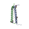
| Entry | Database: PDB / ID: 2kpf | ||||||
|---|---|---|---|---|---|---|---|
| Title | Spatial structure of the dimeric transmembrane domain of glycophorin A in bicelles soluton | ||||||
 Components Components | Glycophorin-A | ||||||
 Keywords Keywords |  MEMBRANE PROTEIN / Glycophorin A / transmembrane dimer / micelles / bicelles / Blood group antigen / Cell membrane / Glycoprotein / Host-virus interaction / Membrane / Sialic acid / Transmembrane MEMBRANE PROTEIN / Glycophorin A / transmembrane dimer / micelles / bicelles / Blood group antigen / Cell membrane / Glycoprotein / Host-virus interaction / Membrane / Sialic acid / Transmembrane | ||||||
| Function / homology |  Function and homology informationankyrin-1 complex / Cell surface interactions at the vascular wall / virus receptor activity / nucleoplasm / membrane / identical protein binding / plasma membrane / cytosol Function and homology informationankyrin-1 complex / Cell surface interactions at the vascular wall / virus receptor activity / nucleoplasm / membrane / identical protein binding / plasma membrane / cytosolSimilarity search - Function | ||||||
| Biological species |  Homo sapiens (human) Homo sapiens (human) | ||||||
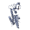
| Method | SOLUTION NMR / torsion angle dynamics | ||||||
| Model details | fewest violations, model 1 | ||||||
 Authors Authors | Mineev, K.S. / Bocharov, E.V. / Goncharuk, M.V. / Arseniev, A.S. / Volynsky, P.E. / Efremov, R.G. | ||||||
 Citation Citation | Journal: Acta Naturae / Year: 2011 Title: Dimeric structure of the transmembrane domain of glycophorin a in lipidic and detergent environments. Authors: Mineev, K.S. / Bocharov, E.V. / Volynsky, P.E. / Goncharuk, M.V. / Tkach, E.N. / Ermolyuk, Y.S. / Schulga, A.A. / Chupin, V.V. / Maslennikov, I.V. / Efremov, R.G. / Arseniev, A.S. | ||||||
| History |
|
- Structure visualization
| Structure viewer | Molecule: MolmilJmol/JSmol |
|---|
- Downloads & links
-Download
| PDBx/mmCIF format | 2kpf.cif.gz | 486.3 KB | Display | PDBx/mmCIF format |
|---|---|---|---|---|
| PDB format | pdb2kpf.ent.gz | 409.5 KB | Display | PDB format |
| PDBx/mmJSON format | 2kpf.json.gz | Tree view | PDBx/mmJSON format | |
| Others |  Other downloads Other downloads |
-Validation report
| Arichive directory | https://data.pdbj.org/pub/pdb/validation_reports/kp/2kpfftp://data.pdbj.org/pub/pdb/validation_reports/kp/2kpf | HTTPS FTP |
|---|
-Related structure data
| Related structure data | 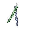 2kpeC C: citing same article ( |
|---|---|
| Similar structure data | |
| Other databases |
|


-Links
 PDBj
PDBj- Assembly
| Deposited unit | 
| |||||||||
|---|---|---|---|---|---|---|---|---|---|---|
| 1 |
| |||||||||
| NMR ensembles |
|
-Components
| #1: Protein/peptide | Mass: 4240.090 Da / Num. of mol.: 2 / Fragment: transmembrane domain (UNP residues 80-117) Source method: isolated from a genetically manipulated source Source: (gene. exp.) Homo sapiens (human) / Gene: GYPA, GPA / Production host:  Escherichia coli (E. coli) / References: UniProt: P02724 Escherichia coli (E. coli) / References: UniProt: P02724 |
|---|
-Experimental details
-Experiment
| Experiment | Method: SOLUTION NMR | ||||||||||||||||||||||||||||||||||||||||
|---|---|---|---|---|---|---|---|---|---|---|---|---|---|---|---|---|---|---|---|---|---|---|---|---|---|---|---|---|---|---|---|---|---|---|---|---|---|---|---|---|---|
| NMR experiment |
|
- Sample preparation
| Details | Contents: 1mM unlabeled Glycophorin A, 1mM 13-C/15-N labeled Glycophorin A, 16mM DMPC d-54, 64 mM DHPC d-22, 95% H2O/5% D2O Solvent system: 95% H2O/5% D2O | ||||||||||||||||
|---|---|---|---|---|---|---|---|---|---|---|---|---|---|---|---|---|---|
| Sample |
| ||||||||||||||||
| Sample conditions | Ionic strength: 0 / pH: 5.5 / Pressure: ambient / Temperature: 313 K |
-NMR measurement
| NMR spectrometer | Type: Varian Unity / Manufacturer: Varian / Model: UNITY / Field strength: 600 MHz |
|---|
- Processing
| NMR software |
| ||||||||||||
|---|---|---|---|---|---|---|---|---|---|---|---|---|---|
| Refinement | Method: torsion angle dynamics / Software ordinal: 1 / Details: CYANA | ||||||||||||
| NMR representative | Selection criteria: fewest violations | ||||||||||||
| NMR ensemble | Conformer selection criteria: target function / Conformers calculated total number: 200 / Conformers submitted total number: 20 |
