 Movie
Movie Controller


+ Open data

- Basic information
| Entry | Database: PDB / ID: 2koj | ||||||
|---|---|---|---|---|---|---|---|
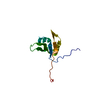
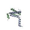
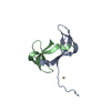
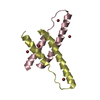
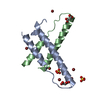
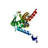
| Title | Solution structure of mouse Par-3 PDZ2 (residues 450-558) | ||||||
 Components Components | Partitioning defective 3 homolog | ||||||
 Keywords Keywords |  SIGNALING PROTEIN / Par-3 / PDZ domain / structural genomics / Alternative splicing / Cell cycle / Cell division / Cell junction / Coiled coil / Cytoplasm / Cytoskeleton / Membrane / Phosphoprotein / Tight junction / PSI-2 / Protein Structure Initiative / Center for Eukaryotic Structural Genomics / CESG SIGNALING PROTEIN / Par-3 / PDZ domain / structural genomics / Alternative splicing / Cell cycle / Cell division / Cell junction / Coiled coil / Cytoplasm / Cytoskeleton / Membrane / Phosphoprotein / Tight junction / PSI-2 / Protein Structure Initiative / Center for Eukaryotic Structural Genomics / CESG | ||||||
| Function / homology |  Function and homology information Function and homology informationTight junction interactions / regulation of actin filament-based process / internode region of axon / regulation of cellular localization / TGF-beta receptor signaling in EMT (epithelial to mesenchymal transition) / apical constriction / establishment of centrosome localization / lateral loop / positive regulation of myelination / establishment of epithelial cell polarity ...Tight junction interactions / regulation of actin filament-based process / internode region of axon / regulation of cellular localization / TGF-beta receptor signaling in EMT (epithelial to mesenchymal transition) / apical constriction / establishment of centrosome localization / lateral loop / positive regulation of myelination / establishment of epithelial cell polarity / Schmidt-Lanterman incisure / bicellular tight junction assembly / myelination in peripheral nervous system / phosphatidylinositol-3-phosphate binding / establishment or maintenance of epithelial cell apical/basal polarity / protein targeting to membrane / wound healing, spreading of cells / centrosome localization / apical junction complex / establishment or maintenance of cell polarity / establishment of cell polarity / negative regulation of peptidyl-threonine phosphorylation / phosphatidylinositol-3,4,5-trisphosphate binding / positive regulation of receptor internalization / bicellular tight junction / endomembrane system / axonal growth cone / phosphatidylinositol-4,5-bisphosphate binding / phosphatidylinositol binding / adherens junction / protein localization / microtubule cytoskeleton organization / cell-cell adhesion / spindle / cell-cell junction / cell junction / cell cortex / protein phosphatase binding / cell adhesion / apical plasma membrane / cell cycle / cell division / neuronal cell body / protein-containing complex / identical protein binding / cytoplasmSimilarity search - Function | ||||||
| Biological species |  Mus musculus (house mouse) Mus musculus (house mouse) | ||||||
| Method | SOLUTION NMR / AUTOMATED METHODS WERE USED FOR BACKBONE CHEMICAL SHIFT ASSIGNMENT, ITERATIVE NOE REFINEMENT. FINAL STRUCTURES WERE OBTAINED BY MOLECULAR DYNAMICS IN EXPLICIT SOLVENT | ||||||
| Model details | lowest energy, model 1 | ||||||
 Authors Authors | Volkman, B.F. / Tyler, R.C. / Peterson, F.C. / Center for Eukaryotic Structural Genomics (CESG) | ||||||
 Citation Citation | Journal: Structural Bioinformatics, 2nd edition / Year: 2009 Title: Macromolecular Structure Determination by NMR Sepectroscopy Authors: Markley, J.L. / Bahrami, A. / Eghbalnia, H.R. / Peterson, F.C. / Ulrich, E.L. / Westler, W.M. / Volkman, B.F. | ||||||
| History |
|
- Structure visualization
| Structure viewer | Molecule: MolmilJmol/JSmol |
|---|
- Downloads & links
-Download
| PDBx/mmCIF format | 2koj.cif.gz | 743.1 KB | Display | PDBx/mmCIF format |
|---|---|---|---|---|
| PDB format | pdb2koj.ent.gz | 627.4 KB | Display | PDB format |
| PDBx/mmJSON format | 2koj.json.gz | Tree view | PDBx/mmJSON format | |
| Others |  Other downloads Other downloads |
-Validation report
| Arichive directory | https://data.pdbj.org/pub/pdb/validation_reports/ko/2kojftp://data.pdbj.org/pub/pdb/validation_reports/ko/2koj | HTTPS FTP |
|---|
-Related structure data
| Similar structure data |
|---|

-Links
 PDBj
PDBj- Assembly
| Deposited unit | 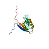
| |||||||||
|---|---|---|---|---|---|---|---|---|---|---|
| 1 |
| |||||||||
| NMR ensembles |
|
-Components
| #1: Protein | Mass: 12044.837 Da / Num. of mol.: 1 / Fragment: PDZ 2 domain Source method: isolated from a genetically manipulated source Source: (gene. exp.) Mus musculus (house mouse) / Gene: Par-3, Par3, Pard3 / Production host:  Escherichia coli (E. coli) / Strain (production host): SG130099[pREP4] / References: UniProt: Q99NH2 Escherichia coli (E. coli) / Strain (production host): SG130099[pREP4] / References: UniProt: Q99NH2 |
|---|
-Experimental details
-Experiment
| Experiment | Method: SOLUTION NMR | ||||||||||||||||
|---|---|---|---|---|---|---|---|---|---|---|---|---|---|---|---|---|---|
| NMR experiment |
|
- Sample preparation
| Details | Contents: 1 mM [U-100% 13C; U-100% 15N] mPar3 PDZ2, 20 mM sodium phosphate, 50 mM sodium chloride, 0.02 % sodium azide, 90% H2O, 10% D2O Solvent system: 90% H2O/10% D2O | ||||||||||||||||||||
|---|---|---|---|---|---|---|---|---|---|---|---|---|---|---|---|---|---|---|---|---|---|
| Sample |
| ||||||||||||||||||||
| Sample conditions | Ionic strength: 54 / pH: 7.0 / Pressure: AMBIENT / Temperature: 298 K |
-NMR measurement
| NMR spectrometer | Type: Bruker Avance II / Manufacturer: Bruker / Model: AVANCE II / Field strength: 600 MHz |
|---|
- Processing
| NMR software |
| ||||||||||||||||||||||||||||||||
|---|---|---|---|---|---|---|---|---|---|---|---|---|---|---|---|---|---|---|---|---|---|---|---|---|---|---|---|---|---|---|---|---|---|
| Refinement | Method: AUTOMATED METHODS WERE USED FOR BACKBONE CHEMICAL SHIFT ASSIGNMENT, ITERATIVE NOE REFINEMENT. FINAL STRUCTURES WERE OBTAINED BY MOLECULAR DYNAMICS IN EXPLICIT SOLVENT Software ordinal: 1 Details: STRUCTURES ARE BASED ON A TOTAL OF 1307 NOE CONSTRAINTS (219 INTRA, 388 SEQUENTIAL, 209 MEDIUM, AND 491 LONG RANGE) AND 117 PHI AND PSI DIHEDRAL ANGLE CONSTRAINTS. | ||||||||||||||||||||||||||||||||
| NMR constraints | NOE constraints total: 1307 / NOE intraresidue total count: 219 / NOE long range total count: 491 / NOE medium range total count: 209 / NOE sequential total count: 388 | ||||||||||||||||||||||||||||||||
| NMR representative | Selection criteria: lowest energy | ||||||||||||||||||||||||||||||||
| NMR ensemble | Conformer selection criteria: target function / Conformers calculated total number: 100 / Conformers submitted total number: 20 |
