 Movie
Movie Controller

[English] 日本語
 Yorodumi
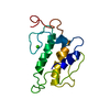
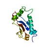
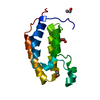
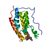
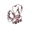
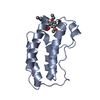
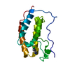
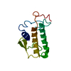
- PDB-2kld: Solution Structure of the Calcium Binding Domain of the C-termina... -
+ Open data

- Basic information
| Entry | Database: PDB / ID: 2kld | ||||||
|---|---|---|---|---|---|---|---|
| Title | Solution Structure of the Calcium Binding Domain of the C-terminal Cytosolic Domain of Polycystin-2 | ||||||
 Components Components | Polycystin-2 Polycystin 2 Polycystin 2 | ||||||
 Keywords Keywords | MEMBRANE PROTEIN / PC2 / PKD2 / Calcium binding domain / EF hand / Cytosolic / Calcium / Coiled coil / Disease mutation / Glycoprotein / Ion transport / Ionic channel / Membrane / Phosphoprotein / Polymorphism / Transmembrane / Transport | ||||||
| Function / homology |  Function and homology information Function and homology informationdetection of nodal flow / metanephric smooth muscle tissue development / metanephric cortex development / metanephric cortical collecting duct development / metanephric distal tubule development / polycystin complex / mesonephric tubule development / mesonephric duct development / : / metanephric part of ureteric bud development ...detection of nodal flow / metanephric smooth muscle tissue development / metanephric cortex development / metanephric cortical collecting duct development / metanephric distal tubule development / polycystin complex / mesonephric tubule development / mesonephric duct development / : / metanephric part of ureteric bud development / determination of liver left/right asymmetry / renal tubule morphogenesis / metanephric ascending thin limb development / HLH domain binding / basal cortex / metanephric mesenchyme development / metanephric S-shaped body morphogenesis / renal artery morphogenesis / positive regulation of inositol 1,4,5-trisphosphate-sensitive calcium-release channel activity / migrasome / cilium organization / VxPx cargo-targeting to cilium / detection of mechanical stimulus / regulation of calcium ion import / cation channel complex / calcium-induced calcium release activity / muscle alpha-actinin binding / placenta blood vessel development / voltage-gated monoatomic ion channel activity / cellular response to hydrostatic pressure / outward rectifier potassium channel activity / voltage-gated monoatomic cation channel activity / non-motile cilium / cellular response to fluid shear stress / cellular response to osmotic stress / voltage-gated sodium channel activity / actinin binding / motile cilium / transcription regulator inhibitor activity / inorganic cation transmembrane transport / determination of left/right symmetry / aorta development / protein heterotetramerization / neural tube development / ciliary membrane / branching involved in ureteric bud morphogenesis / negative regulation of G1/S transition of mitotic cell cycle / spinal cord development / heart looping / cytoplasmic side of endoplasmic reticulum membrane / voltage-gated potassium channel activity / potassium channel activity / cell surface receptor signaling pathway via JAK-STAT / centrosome duplication / sodium ion transmembrane transport / negative regulation of ryanodine-sensitive calcium-release channel activity / voltage-gated calcium channel activity / embryonic placenta development / cellular response to cAMP / release of sequestered calcium ion into cytosol / monoatomic cation channel activity / potassium ion transmembrane transport / cellular response to calcium ion / cytoskeletal protein binding / basal plasma membrane / ciliary basal body / liver development / establishment of localization in cell / lumenal side of endoplasmic reticulum membrane / protein tetramerization / phosphoprotein binding / calcium ion transmembrane transport / cytoplasmic vesicle membrane / cilium / mitotic spindle / Wnt signaling pathway / intracellular calcium ion homeostasis / cellular response to reactive oxygen species / positive regulation of nitric oxide biosynthetic process / calcium ion transport / cell-cell junction / lamellipodium / regulation of cell population proliferation / heart development / ATPase binding / positive regulation of cytosolic calcium ion concentration / basolateral plasma membrane / protein homotetramerization / transmembrane transporter binding / regulation of cell cycle / negative regulation of cell population proliferation / signaling receptor binding / calcium ion binding / endoplasmic reticulum membrane / positive regulation of gene expression / Golgi apparatus / endoplasmic reticulum / protein homodimerization activity / positive regulation of transcription by RNA polymerase II / extracellular exosomeSimilarity search - Function | ||||||
| Biological species |  Homo sapiens (human) Homo sapiens (human) | ||||||
| Method | SOLUTION NMR / restrained molecular dynamics, simulated annealing | ||||||
| Model details | lowest energy, model 1 | ||||||
 Authors Authors | Kalbitzer, H.R. | ||||||
 Citation Citation | Journal: Biomol.Nmr Assign. / Year: 2009 Title: NMR-assignments of a cytosolic domain of the C-terminus of polycystin-2 Authors: Schumann, F.H. / Hoffmeister, H. / Schmidt, M. / Bader, R. / Besl, E. / Witzgall, R. / Kalbitzer, H.R. #1: Journal: J.Biol.Chem. / Year: 2009 Title: Ca2+-dependent conformational changes in a C-terminal cytosolic domain of polycystin-2 Authors: Schumann, F. / Hoffmeister, H. / Bader, R. / Schmidt, M. / Witzgall, R. / Kalbitzer, H.R. | ||||||
| History |
|
- Structure visualization
| Structure viewer | Molecule: MolmilJmol/JSmol |
|---|
- Downloads & links
-Download
| PDBx/mmCIF format | 2kld.cif.gz | 230.6 KB | Display | PDBx/mmCIF format |
|---|---|---|---|---|
| PDB format | pdb2kld.ent.gz | 195.1 KB | Display | PDB format |
| PDBx/mmJSON format | 2kld.json.gz | Tree view | PDBx/mmJSON format | |
| Others |  Other downloads Other downloads |
-Validation report
| Arichive directory | https://data.pdbj.org/pub/pdb/validation_reports/kl/2kldftp://data.pdbj.org/pub/pdb/validation_reports/kl/2kld | HTTPS FTP |
|---|
-Related structure data


-Links
 PDBj
PDBj
- Assembly
| Deposited unit | 
| |||||||||
|---|---|---|---|---|---|---|---|---|---|---|
| 1 |
| |||||||||
| NMR ensembles |
|
-Components
| #1: Protein | Polycystin 2 / Polycystic kidney disease 2 protein / Autosomal dominant polycystic kidney disease type II protein ...Polycystic kidney disease 2 protein / Autosomal dominant polycystic kidney disease type II protein / Polycystwin / R48321 Mass: 14024.415 Da / Num. of mol.: 1 / Fragment: residues 680-796 Source method: isolated from a genetically manipulated source Source: (gene. exp.) Homo sapiens (human) / Gene: PKD2 / Production host:  Escherichia coli (E. coli) / References: UniProt: Q13563 Escherichia coli (E. coli) / References: UniProt: Q13563 |
|---|
-Experimental details
-Experiment
| Experiment | Method: SOLUTION NMR | ||||||||||||||||||||||||||||||||||||||||||||
|---|---|---|---|---|---|---|---|---|---|---|---|---|---|---|---|---|---|---|---|---|---|---|---|---|---|---|---|---|---|---|---|---|---|---|---|---|---|---|---|---|---|---|---|---|---|
| NMR experiment |
|
- Sample preparation
| Details | Contents: 0.5mM [U-100% 13C; U-100% 15N] Polycystin-2 Polypeptide-1, 5mM Ca2+-2, 0.1mM DSS-3, 10mM potassium phosphate buffer-4, 500mM NaCl-5, 2mM DTE-6, 90% H2O/10% D2O Solvent system: 90% H2O/10% D2O | ||||||||||||||||||||||||||||
|---|---|---|---|---|---|---|---|---|---|---|---|---|---|---|---|---|---|---|---|---|---|---|---|---|---|---|---|---|---|
| Sample |
| ||||||||||||||||||||||||||||
| Sample conditions | Ionic strength: 0.51 / pH: 6.8 / Pressure: ambient / Temperature: 293 K |
-NMR measurement
| NMR spectrometer |
|
|---|
- Processing
| NMR software |
| ||||||||||||||||||||
|---|---|---|---|---|---|---|---|---|---|---|---|---|---|---|---|---|---|---|---|---|---|
| Refinement | Method: restrained molecular dynamics, simulated annealing / Software ordinal: 1 Details: THE STRUCTURES WERE REFINED IN EXPLICIT WATER ; Linge, J.P., Williams, M.A., Spronk, C.A.E.M., Bonvin, A.M.J.J. & Nilges, M. (2003). Refinement of protein structures in explicit solvent. ...Details: THE STRUCTURES WERE REFINED IN EXPLICIT WATER ; Linge, J.P., Williams, M.A., Spronk, C.A.E.M., Bonvin, A.M.J.J. & Nilges, M. (2003). Refinement of protein structures in explicit solvent. Proteins: Struct. Funct. Genet. 50, 496-506 | ||||||||||||||||||||
| NMR representative | Selection criteria: lowest energy | ||||||||||||||||||||
| NMR ensemble | Conformer selection criteria: structures with the lowest energy Conformers calculated total number: 1000 / Conformers submitted total number: 10 / Representative conformer: 1 |
