 Movie
Movie Controller

[English] 日本語
 Yorodumi
- PDB-2kb7: Hybrid solution and solid-state NMR structure of monomeric phosph... -
+ Open data

- Basic information
| Entry | Database: PDB / ID: 2kb7 | ||||||
|---|---|---|---|---|---|---|---|
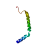
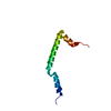
| Title | Hybrid solution and solid-state NMR structure of monomeric phospholamban in lipid bilayers | ||||||
 Components Components | Phospholamban | ||||||
 Keywords Keywords | MEMBRANE PROTEIN / phospholamban / PISEMA / hybrid method / lipid bilayers / topology | ||||||
| Function / homology |  Function and homology information Function and homology informationnegative regulation of calcium ion binding / negative regulation of calcium ion import into sarcoplasmic reticulum / negative regulation of ATPase-coupled calcium transmembrane transporter activity / adenylate cyclase-activating adrenergic receptor signaling pathway involved in heart process / regulation of relaxation of muscle / regulation of the force of heart contraction by cardiac conduction / calcium ion-transporting ATPase complex / negative regulation of calcium ion transmembrane transporter activity / acrosome assembly / negative regulation of calcium ion import ...negative regulation of calcium ion binding / negative regulation of calcium ion import into sarcoplasmic reticulum / negative regulation of ATPase-coupled calcium transmembrane transporter activity / adenylate cyclase-activating adrenergic receptor signaling pathway involved in heart process / regulation of relaxation of muscle / regulation of the force of heart contraction by cardiac conduction / calcium ion-transporting ATPase complex / negative regulation of calcium ion transmembrane transporter activity / acrosome assembly / negative regulation of calcium ion import / negative regulation of catalytic activity / ATPase inhibitor activity / cardiac muscle tissue development / regulation of cardiac muscle cell contraction / enzyme inhibitor activity / negative regulation of heart rate / muscle cell cellular homeostasis / regulation of calcium ion transport / regulation of cardiac muscle contraction by regulation of the release of sequestered calcium ion / Notch signaling pathway / sarcoplasmic reticulum membrane / mitochondrial membrane / intracellular calcium ion homeostasis / ATPase binding / endoplasmic reticulum membrane / perinuclear region of cytoplasm / endoplasmic reticulum / protein homodimerization activity / identical protein bindingSimilarity search - Function | ||||||
| Biological species |  Escherichia coli (E. coli) Escherichia coli (E. coli) | ||||||
| Method | SOLUTION NMR / SOLID-STATE NMR / simulated annealing | ||||||
 Authors Authors | Traaseth, N.J. / Shi, L. / Verardi, R. / Veglia, G. | ||||||
 Citation Citation | Journal: Proc.Natl.Acad.Sci.USA / Year: 2009 Title: Structure and topology of monomeric phospholamban in lipid membranes determined by a hybrid solution and solid-state NMR approach. Authors: Traaseth, N.J. / Shi, L. / Verardi, R. / Mullen, D.G. / Barany, G. / Veglia, G. #1: Journal: J.Biomol.Nmr / Year: 2009 Title: A refinement protocol to determine structure, topology, and depth of insertion of membrane proteins using hybrid solution and solid-state NMR restraints. Authors: Shi, L. / Traaseth, N.J. / Verardi, R. / Cembran, A. / Gao, J. / Veglia, G. | ||||||
| History |
|
- Structure visualization
| Structure viewer | Molecule: MolmilJmol/JSmol |
|---|
- Downloads & links
-Download
| PDBx/mmCIF format | 2kb7.cif.gz | 355.3 KB | Display | PDBx/mmCIF format |
|---|---|---|---|---|
| PDB format | pdb2kb7.ent.gz | 299.7 KB | Display | PDB format |
| PDBx/mmJSON format | 2kb7.json.gz | Tree view | PDBx/mmJSON format | |
| Others |  Other downloads Other downloads |
-Validation report
| Arichive directory | https://data.pdbj.org/pub/pdb/validation_reports/kb/2kb7ftp://data.pdbj.org/pub/pdb/validation_reports/kb/2kb7 | HTTPS FTP |
|---|
-Related structure data
| Related structure data | |
|---|---|
| Similar structure data |


-Links
 PDBj
PDBj- Assembly
| Deposited unit | 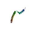
| |||||||||
|---|---|---|---|---|---|---|---|---|---|---|
| 1 |
| |||||||||
| NMR ensembles |
|
-Components
| #1: Protein | Mass: 6150.477 Da / Num. of mol.: 1 Source method: isolated from a genetically manipulated source Source: (gene. exp.) Escherichia coli (E. coli) / Strain: Bl21de3phi / Gene: pln / Plasmid: pMAL / Production host: Escherichia coli (E. coli) / References: UniProt: P61015*PLUS |
|---|
-Experimental details
-Experiment
| Experiment |
| ||||||||||||||||||||||||||||
|---|---|---|---|---|---|---|---|---|---|---|---|---|---|---|---|---|---|---|---|---|---|---|---|---|---|---|---|---|---|
| NMR experiment |
|
- Sample preparation
| Details |
| |||||||||||||||
|---|---|---|---|---|---|---|---|---|---|---|---|---|---|---|---|---|
| Sample |
| |||||||||||||||
| Sample conditions | pH: 6.0 / Pressure: ambient / Temperature units: K |
-Data collection
| NMR spectrometer |
|
|---|
- Processing
| NMR software |
| ||||||||||||||||
|---|---|---|---|---|---|---|---|---|---|---|---|---|---|---|---|---|---|
| Refinement | Method: simulated annealing / Software ordinal: 1 Details: Authors state that they define the z-axis to be parallel with the bilayer normal. Also, the origin (or in other words, when the z Cartesian coordinate is 0) is defined to be the center of the lipid bilayer. | ||||||||||||||||
| NMR representative | Selection criteria: fewest violations | ||||||||||||||||
| NMR ensemble | Conformer selection criteria: structures with the least restraint violations Conformers calculated total number: 200 / Conformers submitted total number: 20 |
