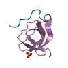
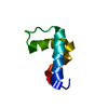
[English] 日本語
Yorodumi
- PDB-2jku: Crystal structure of the N-terminal region of the biotin acceptor... -

+
Open data


ID or keywords:

Loading...

-
Basic information

Entry
Database: PDB / ID: 2jku
TitleCrystal structure of the N-terminal region of the biotin acceptor domain of human propionyl-CoA carboxylase
ComponentsPROPIONYL-COA CARBOXYLASE ALPHA CHAIN, MITOCHONDRIAL
KeywordsLIGASE / BIOTIN / ATP-BINDING / DISEASE MUTATION / NUCLEOTIDE-BINDING / MITOCHONDRION / PHOSPHOPROTEIN / TRANSIT PEPTIDE
Function / homology
Function and homology information


short-chain fatty acid catabolic process / branched-chain amino acid metabolic process / Propionyl-CoA catabolism / propionyl-CoA carboxylase / propionyl-CoA carboxylase activity / Defective HLCS causes multiple carboxylase deficiency / Biotin transport and metabolism / biotin binding / catalytic complex / fatty acid metabolic process ...short-chain fatty acid catabolic process / branched-chain amino acid metabolic process / Propionyl-CoA catabolism / propionyl-CoA carboxylase / propionyl-CoA carboxylase activity / Defective HLCS causes multiple carboxylase deficiency / Biotin transport and metabolism / biotin binding / catalytic complex / fatty acid metabolic process / mitochondrial matrix / enzyme binding / mitochondrion / ATP binding / metal ion binding / cytosol
Similarity search - Function
Propionyl-coenzyme A carboxylase, BT domain / Propionyl-coenzyme A carboxylase BT domain / Biotin-binding site / Biotin-requiring enzymes attachment site. / Biotin carboxylase-like, N-terminal domain / Biotin carboxylase, C-terminal / Biotin carboxylation domain / Biotin carboxylase, N-terminal domain / Biotin carboxylase C-terminal domain / Biotin carboxylation domain profile. ...Propionyl-coenzyme A carboxylase, BT domain / Propionyl-coenzyme A carboxylase BT domain / Biotin-binding site / Biotin-requiring enzymes attachment site. / Biotin carboxylase-like, N-terminal domain / Biotin carboxylase, C-terminal / Biotin carboxylation domain / Biotin carboxylase, N-terminal domain / Biotin carboxylase C-terminal domain / Biotin carboxylation domain profile. / Biotin carboxylase C-terminal domain / Carbamoyl-phosphate synthase subdomain signature 1. / Carbamoyl-phosphate synthetase large subunit-like, ATP-binding domain / Carbamoyl-phosphate synthase L chain, ATP binding domain / Biotin-requiring enzyme / Rudiment single hybrid motif / Biotinyl/lipoyl domain profile. / Biotin/lipoyl attachment / Single hybrid motif / ATP-grasp fold, subdomain 1 / Pre-ATP-grasp domain superfamily / ATP-grasp fold / ATP-grasp fold profile. / Carbamoyl-phosphate synthase subdomain signature 2.
Similarity search - Domain/homology
Propionyl-CoA carboxylase alpha chain, mitochondrial
Similarity search - Component
Biological speciesHOMO SAPIENS (human)
MethodX-RAY DIFFRACTION / SYNCHROTRON / MOLECULAR REPLACEMENT / Resolution: 1.5 Å
AuthorsHealy, S. / Yue, W.W. / Kochan, G. / Pilka, E.S. / Murray, J.W. / Roos, A.K. / Filippakopoulos, P. / von Delft, F. / Arrowsmith, C. / Wikstrom, M. ...Healy, S. / Yue, W.W. / Kochan, G. / Pilka, E.S. / Murray, J.W. / Roos, A.K. / Filippakopoulos, P. / von Delft, F. / Arrowsmith, C. / Wikstrom, M. / Edwards, A. / Bountra, C. / Gravel, R.A. / Oppermann, U.
CitationJournal: Biochemistry / Year: 2010
Title: Structural impact of human and Escherichia coli biotin carboxyl carrier proteins on biotin attachment.
Authors: Healy, S. / McDonald, M.K. / Wu, X. / Yue, W.W. / Kochan, G. / Oppermann, U. / Gravel, R.A.
History
DepositionAug 30, 2008Deposition site: PDBE / Processing site: PDBE
Revision 1.0Sep 9, 2008Provider: repository / Type: Initial release
Revision 1.1Dec 4, 2013Group: Source and taxonomy / Version format compliance
Revision 1.2Feb 28, 2018Group: Database references / Category: citation / citation_author
Item: _citation.journal_id_ISSN / _citation.page_last ..._citation.journal_id_ISSN / _citation.page_last / _citation.pdbx_database_id_DOI / _citation.title / _citation_author.name
Revision 1.3Mar 4, 2020Group: Derived calculations / Other
Category: pdbx_database_status / pdbx_struct_assembly ...pdbx_database_status / pdbx_struct_assembly / pdbx_struct_assembly_gen / pdbx_struct_assembly_prop / pdbx_struct_oper_list
Item: _pdbx_database_status.status_code_sf / _pdbx_struct_assembly.details ..._pdbx_database_status.status_code_sf / _pdbx_struct_assembly.details / _pdbx_struct_assembly.oligomeric_count / _pdbx_struct_assembly.oligomeric_details / _pdbx_struct_assembly_gen.oper_expression
Revision 1.4Dec 13, 2023Group: Data collection / Database references ...Data collection / Database references / Derived calculations / Refinement description
Category: chem_comp_atom / chem_comp_bond ...chem_comp_atom / chem_comp_bond / database_2 / pdbx_initial_refinement_model / struct_site
Item: _database_2.pdbx_DOI / _database_2.pdbx_database_accession ..._database_2.pdbx_DOI / _database_2.pdbx_database_accession / _struct_site.pdbx_auth_asym_id / _struct_site.pdbx_auth_comp_id / _struct_site.pdbx_auth_seq_id

-
Structure visualization

Structure viewerMolecule:
MolmilJmol/JSmol

Downloads & links

-
Assembly

Deposited unit
A: PROPIONYL-COA CARBOXYLASE ALPHA CHAIN, MITOCHONDRIAL
hetero molecules


Theoretical massNumber of molelcules
Total (without water)10,3293
Polymers9,9401
Non-polymers3882
Water1,04558
1
A: PROPIONYL-COA CARBOXYLASE ALPHA CHAIN, MITOCHONDRIAL
hetero molecules

A: PROPIONYL-COA CARBOXYLASE ALPHA CHAIN, MITOCHONDRIAL
hetero molecules


Theoretical massNumber of molelcules
Total (without water)20,6576
Polymers19,8802
Non-polymers7774
Water362
TypeNameSymmetry operationNumber
identity operation1_555x,y,z1
crystal symmetry operation11_455-x+y-1,y,-z1
Buried area3720 Å2
ΔGint-13 kcal/mol
Surface area4230 Å2
MethodPISA
Unit cell
Length a, b, c (Å)56.107, 56.107, 58.083
Angle α, β, γ (deg.)90.00, 90.00, 120.00
Int Tables number181
Space group name H-MP6422
Components on special symmetry positions
IDModelComponents
11A-2002-

HOH

21A-2048-

HOH

-
Components

#1: Protein PROPIONYL-COA CARBOXYLASE ALPHA CHAIN, MITOCHONDRIAL / PROPANOYL-COA\:CARBON DIOXIDE LIGASE SUBUNIT ALPHA


Mass: 9940.248 Da / Num. of mol.: 1 / Fragment: BIOTIN ACCEPTOR DOMAIN, RESIDUES 633-703
Source method: isolated from a genetically manipulated source
Source: (gene. exp.) HOMO SAPIENS (human) / Organelle: MITOCHONDRIAMitochondrion / Plasmid: PNIC28-BSA4 / Production host: ESCHERICHIA COLI (E. coli) / Strain (production host): BL21(DE3) / Variant (production host): R3-PRARE2 / References: UniProt: P05165, propionyl-CoA carboxylase
#2: Chemical ChemComp-PG4 / TETRAETHYLENE GLYCOL / Polyethylene glycol


Mass: 194.226 Da / Num. of mol.: 2 / Source method: obtained synthetically / Formula: C8H18O5 / Comment: precipitant*YM
#3: Water ChemComp-HOH / water / Water


Mass: 18.015 Da / Num. of mol.: 58 / Source method: isolated from a natural source / Formula: H2O

-
Experimental details

-
Experiment

ExperimentMethod: X-RAY DIFFRACTION / Number of used crystals: 1

-
Sample preparation

CrystalDensity Matthews: 3.57 Å3/Da / Density % sol: 65.53 % / Description: NONE
Crystal growDetails: 20% PEG 3350, 0.20M NA FORMATE

-
Data collection

DiffractionMean temperature: 100 K
Diffraction sourceSource: SYNCHROTRON / Site: SLS / Beamline: X10SA / Wavelength: 0.97628
DetectorType: MARRESEARCH / Detector: CCD / Date: Jun 21, 2008
RadiationMonochromator: SI (111) / Protocol: SINGLE WAVELENGTH / Monochromatic (M) / Laue (L): M / Scattering type: x-ray
Radiation wavelengthWavelength: 0.97628 Å / Relative weight: 1
ReflectionResolution: 1.5→48.56 Å / Num. obs: 10251 / % possible obs: 99.9 % / Observed criterion σ(I): 2 / Redundancy: 12.7 % / Rmerge(I) obs: 0.08 / Net I/σ(I): 23.6
Reflection shellResolution: 1.5→1.6 Å / Redundancy: 13.4 % / Rmerge(I) obs: 0.44 / Mean I/σ(I) obs: 5.2 / % possible all: 100

-
Processing

Software
NameVersionClassification
REFMAC5.5.0036refinement
MOSFLMdata reduction
SCALAdata scaling
PHASERphasing
RefinementMethod to determine structure: MOLECULAR REPLACEMENT
Starting model: PDB ENTRIES 2D5D, 3BG3
Resolution: 1.5→28.12 Å / Cor.coef. Fo:Fc: 0.969 / Cor.coef. Fo:Fc free: 0.966 / SU B: 1.564 / SU ML: 0.027 / Cross valid method: THROUGHOUT / ESU R: 0.056 / ESU R Free: 0.05 / Stereochemistry target values: MAXIMUM LIKELIHOOD / Details: HYDROGENS HAVE BEEN ADDED IN THE RIDING POSITIONS.
RfactorNum. reflection% reflectionSelection details
Rfree0.166 438 4.8 %RANDOM
Rwork0.148 ---
obs0.149 8711 100 %-
Solvent computationIon probe radii: 0.8 Å / Shrinkage radii: 0.8 Å / VDW probe radii: 1.2 Å / Solvent model: MASK
Displacement parametersBiso mean: 12.85 Å2
Baniso -1Baniso -2Baniso -3
1--0.06 Å2-0.03 Å20 Å2
2---0.06 Å20 Å2
3---0.1 Å2
Refinement stepCycle: LAST / Resolution: 1.5→28.12 Å
ProteinNucleic acidLigandSolventTotal
Num. atoms242 0 23 58 323
Refine LS restraints
Refine-IDTypeDev idealDev ideal targetNumber
X-RAY DIFFRACTIONr_bond_refined_d0.0110.023289
X-RAY DIFFRACTIONr_bond_other_d0.0020.02202
X-RAY DIFFRACTIONr_angle_refined_deg1.5462.12389
X-RAY DIFFRACTIONr_angle_other_deg0.8933.018469
X-RAY DIFFRACTIONr_dihedral_angle_1_deg5.668541
X-RAY DIFFRACTIONr_dihedral_angle_2_deg25.09526.6676
X-RAY DIFFRACTIONr_dihedral_angle_3_deg10.221547
X-RAY DIFFRACTIONr_dihedral_angle_4_deg10.952151
X-RAY DIFFRACTIONr_chiral_restr0.0770.250
X-RAY DIFFRACTIONr_gen_planes_refined0.0050.021294
X-RAY DIFFRACTIONr_gen_planes_other0.0010.0239
X-RAY DIFFRACTIONr_nbd_refined
X-RAY DIFFRACTIONr_nbd_other
X-RAY DIFFRACTIONr_nbtor_refined
X-RAY DIFFRACTIONr_nbtor_other
X-RAY DIFFRACTIONr_xyhbond_nbd_refined
X-RAY DIFFRACTIONr_xyhbond_nbd_other
X-RAY DIFFRACTIONr_metal_ion_refined
X-RAY DIFFRACTIONr_metal_ion_other
X-RAY DIFFRACTIONr_symmetry_vdw_refined
X-RAY DIFFRACTIONr_symmetry_vdw_other
X-RAY DIFFRACTIONr_symmetry_hbond_refined
X-RAY DIFFRACTIONr_symmetry_hbond_other
X-RAY DIFFRACTIONr_symmetry_metal_ion_refined
X-RAY DIFFRACTIONr_symmetry_metal_ion_other
X-RAY DIFFRACTIONr_mcbond_it2.333188
X-RAY DIFFRACTIONr_mcbond_other
X-RAY DIFFRACTIONr_mcangle_it3.8235311
X-RAY DIFFRACTIONr_mcangle_other
X-RAY DIFFRACTIONr_scbond_it5.8427101
X-RAY DIFFRACTIONr_scbond_other
X-RAY DIFFRACTIONr_scangle_it7.2481175
X-RAY DIFFRACTIONr_scangle_other
X-RAY DIFFRACTIONr_long_range_B_refined
X-RAY DIFFRACTIONr_long_range_B_other
X-RAY DIFFRACTIONr_rigid_bond_restr
X-RAY DIFFRACTIONr_sphericity_free
X-RAY DIFFRACTIONr_sphericity_bonded
LS refinement shellResolution: 1.5→1.54 Å / Total num. of bins used: 20 /
RfactorNum. reflection
Rfree0.224 33
Rwork0.191 614

+
About Yorodumi

-
News

-
Feb 9, 2022. New format data for meta-information of EMDB entries

New format data for meta-information of EMDB entries

  • Version 3 of the EMDB header file is now the official format.
  • The previous official version 1.9 will be removed from the archive.

Related info.:EMDB header

External links:wwPDB to switch to version 3 of the EMDB data model

-
Aug 12, 2020. Covid-19 info

Covid-19 info

URL: https://pdbj.org/emnavi/covid19.php

New page: Covid-19 featured information page in EM Navigator.

Related info.:Covid-19 info / Mar 5, 2020. Novel coronavirus structure data

+
Mar 5, 2020. Novel coronavirus structure data

Novel coronavirus structure data

Related info.:Yorodumi Speices / Aug 12, 2020. Covid-19 info

External links:COVID-19 featured content - PDBj / Molecule of the Month (242):Coronavirus Proteases

+
Jan 31, 2019. EMDB accession codes are about to change! (news from PDBe EMDB page)

EMDB accession codes are about to change! (news from PDBe EMDB page)

  • The allocation of 4 digits for EMDB accession codes will soon come to an end. Whilst these codes will remain in use, new EMDB accession codes will include an additional digit and will expand incrementally as the available range of codes is exhausted. The current 4-digit format prefixed with “EMD-” (i.e. EMD-XXXX) will advance to a 5-digit format (i.e. EMD-XXXXX), and so on. It is currently estimated that the 4-digit codes will be depleted around Spring 2019, at which point the 5-digit format will come into force.
  • The EM Navigator/Yorodumi systems omit the EMD- prefix.

Related info.:Q: What is EMD? / ID/Accession-code notation in Yorodumi/EM Navigator

External links:EMDB Accession Codes are Changing Soon! / Contact to PDBj

+
Jul 12, 2017. Major update of PDB

Major update of PDB

  • wwPDB released updated PDB data conforming to the new PDBx/mmCIF dictionary.
  • This is a major update changing the version number from 4 to 5, and with Remediation, in which all the entries are updated.
  • In this update, many items about electron microscopy experimental information are reorganized (e.g. em_software).
  • Now, EM Navigator and Yorodumi are based on the updated data.

External links:wwPDB Remediation / Enriched Model Files Conforming to OneDep Data Standards Now Available in the PDB FTP Archive

-
Yorodumi

Thousand views of thousand structures

  • Yorodumi is a browser for structure data from EMDB, PDB, SASBDB, etc.
  • This page is also the successor to EM Navigator detail page, and also detail information page/front-end page for Omokage search.
  • The word "yorodu" (or yorozu) is an old Japanese word meaning "ten thousand". "mi" (miru) is to see.

Related info.:EMDB / PDB / SASBDB / Comparison of 3 databanks / Yorodumi Search / Aug 31, 2016. New EM Navigator & Yorodumi / Yorodumi Papers / Jmol/JSmol / Function and homology information / Changes in new EM Navigator and Yorodumi

Read more